 Movie
Movie Controller


+ Open data

- Basic information
| Entry | Database: PDB / ID: 2uwi | ||||||
|---|---|---|---|---|---|---|---|
| Title | Structure of CrmE, a poxvirus TNF receptor | ||||||
 Components Components | CRME PROTEIN | ||||||
 Keywords Keywords |  RECEPTOR / POXVIRUS TNF RECEPTOR / RECEPTOR IMMUNOMODULATOR / TNF ALPHA RECEPTOR RECEPTOR / POXVIRUS TNF RECEPTOR / RECEPTOR IMMUNOMODULATOR / TNF ALPHA RECEPTOR | ||||||
| Function / homology |  Function and homology informationTumor necrosis factor receptor, N-terminal, viral / Tumor Necrosis Factor Receptor, subunit A, domain 2 / Tumor Necrosis Factor Receptor, subunit A; domain 2 / TNFR/NGFR family cysteine-rich region domain profile. / TNFR/NGFR cysteine-rich region / TNFR/NGFR family cysteine-rich region signature. / Tumor necrosis factor receptor / nerve growth factor receptor repeats. / TNFR/NGFR cysteine-rich region / Ribbon / Mainly Beta Function and homology informationTumor necrosis factor receptor, N-terminal, viral / Tumor Necrosis Factor Receptor, subunit A, domain 2 / Tumor Necrosis Factor Receptor, subunit A; domain 2 / TNFR/NGFR family cysteine-rich region domain profile. / TNFR/NGFR cysteine-rich region / TNFR/NGFR family cysteine-rich region signature. / Tumor necrosis factor receptor / nerve growth factor receptor repeats. / TNFR/NGFR cysteine-rich region / Ribbon / Mainly BetaSimilarity search - Domain/homology | ||||||
| Biological species |   VACCINIA VIRUS VACCINIA VIRUS | ||||||
| Method | X-RAY DIFFRACTION / SYNCHROTRON / MAD / Resolution: 2 Å | ||||||
 Authors Authors | Graham, S.C. / Bahar, M.W. / Abrescia, N.G. / Smith, G.L. / Stuart, D.I. / Grimes, J.M. | ||||||
 Citation Citation | Journal: J.Mol.Biol. / Year: 2007 Title: Structure of Crme, a Virus-Encoded Tumour Necrosis Factor Receptor. Authors: Graham, S.C. / Bahar, M.W. / Abrescia, N.G. / Smith, G.L. / Stuart, D.I. / Grimes, J.M. | ||||||
| History |
|
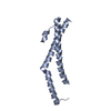
- Structure visualization
| Structure viewer | Molecule: MolmilJmol/JSmol |
|---|
- Downloads & links
-Download
| PDBx/mmCIF format | 2uwi.cif.gz | 59.8 KB | Display | PDBx/mmCIF format |
|---|---|---|---|---|
| PDB format | pdb2uwi.ent.gz | 47.9 KB | Display | PDB format |
| PDBx/mmJSON format | 2uwi.json.gz | Tree view | PDBx/mmJSON format | |
| Others |  Other downloads Other downloads |
-Validation report
| Arichive directory | https://data.pdbj.org/pub/pdb/validation_reports/uw/2uwiftp://data.pdbj.org/pub/pdb/validation_reports/uw/2uwi | HTTPS FTP |
|---|
-Related structure data
| Similar structure data |
|---|


-Links
 PDBj
PDBj
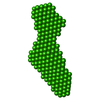
- Assembly
| Deposited unit | 
| ||||||||
|---|---|---|---|---|---|---|---|---|---|
| 1 | 
| ||||||||
| 2 | 
| ||||||||
| Unit cell |
|
-Components
| #1: Protein | Mass: 15872.030 Da / Num. of mol.: 2 / Fragment: CYSTEINE-RICH DOMAINS 1-3, RESIDUES 22-153 Source method: isolated from a genetically manipulated source Source: (gene. exp.) VACCINIA VIRUS / Strain: LISTER / Plasmid: PDEST14CRME / Production host:  Escherichia coli BL21(DE3) (bacteria) / Variant (production host): Rosetta pLySS / References: UniProt: Q8UYL3 Escherichia coli BL21(DE3) (bacteria) / Variant (production host): Rosetta pLySS / References: UniProt: Q8UYL3#2: Water | ChemComp-HOH / | Water Mass: 18.015 Da / Num. of mol.: 192 / Source method: isolated from a natural source / Formula: H2O Mass: 18.015 Da / Num. of mol.: 192 / Source method: isolated from a natural source / Formula: H2OSequence details | CRDS 1-3 OF CRME, C-TERMINAL HIS TAGGED CONSTRUCT. | |
|---|
-Experimental details
-Experiment
| Experiment | Method: X-RAY DIFFRACTION / Number of used crystals: 1 |
|---|
- Sample preparation
| Crystal | Density Matthews: 2.27 Å3/Da / Density % sol: 45.86 % / Description: NONE |
|---|---|
| Crystal grow | Temperature: 293 K / Method: vapor diffusion, sitting drop / pH: 5 Details: 100 NL PROTEIN (6 MG/ML CRME IN 20 MM TRIS 150 MM NACL PH 8.0) AND 100 NL RESERVOIR (0.1 M CITRATE PH 5.0 PLUS 30% W/V PEG 6000) SITTING DROPS AT 20C EQUILIBRATED AGAINST 100 UL RESERVOIRS. |
-Data collection
| Diffraction | Mean temperature: 100 K |
|---|---|
| Diffraction source | Source: SYNCHROTRON / Site: ESRF  / Beamline: ID23-1 / Wavelength: 1.0723 / Beamline: ID23-1 / Wavelength: 1.0723 |
| Detector | Type: ADSC CCD / Detector: CCD / Date: May 14, 2006 / Details: SILICON TOROIDAL MIRROR COATED WITH RHODIUM |
| Radiation | Monochromator: SILICON (1 1 1) CHANNEL- CUT / Protocol: SINGLE WAVELENGTH / Monochromatic (M) / Laue (L): M / Scattering type: x-ray |
| Radiation wavelength | Wavelength: 1.0723 Å / Relative weight: 1 |
| Reflection | Resolution: 2→100 Å / Num. obs: 20089 / % possible obs: 99.9 % / Observed criterion σ(I): -3 / Redundancy: 3.5 % / Rmerge(I) obs: 0.11 / Net I/σ(I): 11.1 |
| Reflection shell | Resolution: 2→2.07 Å / Redundancy: 3.5 % / Rmerge(I) obs: 0.88 / Mean I/σ(I) obs: 1.4 / % possible all: 100 |
- Processing
| Software |
| ||||||||||||||||||||||||||||||||||||||
|---|---|---|---|---|---|---|---|---|---|---|---|---|---|---|---|---|---|---|---|---|---|---|---|---|---|---|---|---|---|---|---|---|---|---|---|---|---|---|---|
| Refinement | Method to determine structure: MAD Starting model: NONE Resolution: 2→39.193 Å / Cross valid method: THROUGHOUT / σ(F): 0 Details: REFINED IN BUSTER TNT BETA VERSION 2.1.1 SIDECHAINS OF RESIDUES A 24 (GLN), A52 (LYS), A53 (TYR), A148 (ARG), B24 (GLN), B86 (ASN), B148 (ARG) WERE NOT VISIBLE IN ELECTRON DENSITY AND WERE NOT MODELLED.
| ||||||||||||||||||||||||||||||||||||||
| Refinement step | Cycle: LAST / Resolution: 2→39.193 Å
| ||||||||||||||||||||||||||||||||||||||
| Refine LS restraints |
|
