 Movie
Movie Controller


+ Open data

- Basic information
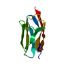
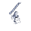
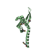
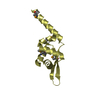
| Entry | Database: PDB / ID: 2rq8 | ||||||
|---|---|---|---|---|---|---|---|
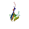
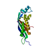
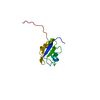
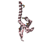
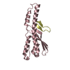
| Title | Solution NMR structure of titin I27 domain mutant | ||||||
 Components Components | Titin | ||||||
 Keywords Keywords | TRANSFERASE / Beta-sandwich / Immunoglobulin-like domain / Alternative splicing / ATP-binding / Calcium / Calmodulin-binding / Cardiomyopathy / Coiled coil / Cytoplasm / Disease mutation / Disulfide bond / Immunoglobulin domain / Isopeptide bond / Kelch repeat / Kinase / Limb-girdle muscular dystrophy / Magnesium / Metal-binding / Nucleotide-binding / Nucleus / Phosphoprotein / Polymorphism / Serine/threonine-protein kinase / TPR repeat / Ubl conjugation / WD repeat | ||||||
| Function / homology |  Function and homology information Function and homology informationsarcomerogenesis / structural molecule activity conferring elasticity / telethonin binding / skeletal muscle myosin thick filament assembly / cardiac myofibril assembly / muscle alpha-actinin binding / detection of muscle stretch / cardiac muscle tissue morphogenesis / regulation of catalytic activity / cardiac muscle hypertrophy ...sarcomerogenesis / structural molecule activity conferring elasticity / telethonin binding / skeletal muscle myosin thick filament assembly / cardiac myofibril assembly / muscle alpha-actinin binding / detection of muscle stretch / cardiac muscle tissue morphogenesis / regulation of catalytic activity / cardiac muscle hypertrophy / mitotic chromosome condensation / Striated Muscle Contraction / M band / actinin binding / I band / cardiac muscle cell development / regulation of protein kinase activity / sarcomere organization / structural constituent of muscle / skeletal muscle thin filament assembly / striated muscle thin filament / striated muscle contraction / protein kinase A signaling / cardiac muscle contraction / muscle contraction / condensed nuclear chromosome / positive regulation of protein secretion / Z disc / response to calcium ion / : / actin filament binding / Platelet degranulation / protein tyrosine kinase activity / protease binding / calmodulin binding / non-specific serine/threonine protein kinase / phosphorylation / protein serine kinase activity / protein serine/threonine kinase activity / calcium ion binding / positive regulation of gene expression / protein kinase binding / enzyme binding / extracellular exosome / extracellular region / ATP binding / identical protein binding / cytosolSimilarity search - Function | ||||||
| Biological species |  Homo sapiens (human) Homo sapiens (human) | ||||||
| Method | SOLUTION NMR / torsion angle dynamics | ||||||
| Model details | lowest energy, model 1 | ||||||
 Authors Authors | Yagawa, K. / Oguro, T. / Momose, T. / Kawano, S. / Sato, T. / Endo, T. | ||||||
 Citation Citation | Journal: Protein Sci. / Year: 2010 Title: Structural basis for unfolding pathway-dependent stability of proteins: Vectorial unfolding vs. global unfolding Authors: Yagawa, K. / Yamano, K. / Oguro, T. / Maeda, M. / Sato, T. / Momose, T. / Kawano, S. / Endo, T. | ||||||
| History |
|
- Structure visualization
| Structure viewer | Molecule: MolmilJmol/JSmol |
|---|
- Downloads & links
-Download
| PDBx/mmCIF format | 2rq8.cif.gz | 584.5 KB | Display | PDBx/mmCIF format |
|---|---|---|---|---|
| PDB format | pdb2rq8.ent.gz | 510.8 KB | Display | PDB format |
| PDBx/mmJSON format | 2rq8.json.gz | Tree view | PDBx/mmJSON format | |
| Others |  Other downloads Other downloads |
-Validation report
| Arichive directory | https://data.pdbj.org/pub/pdb/validation_reports/rq/2rq8ftp://data.pdbj.org/pub/pdb/validation_reports/rq/2rq8 | HTTPS FTP |
|---|
-Related structure data
| Related structure data | |
|---|---|
| Similar structure data |

-Links
 PDBj
PDBj


- Assembly
| Deposited unit | 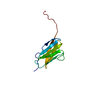
| |||||||||
|---|---|---|---|---|---|---|---|---|---|---|
| 1 |
| |||||||||
| NMR ensembles |
|
-Components
| #1: Protein | / Connectin / Rhabdomyosarcoma antigen MU-RMS-40.14 Mass: 10930.482 Da / Num. of mol.: 1 / Fragment: UNP residues 12677-12765 / Mutation: K3E, Y9P, T42A, A78T Source method: isolated from a genetically manipulated source Source: (gene. exp.) Homo sapiens (human) / Gene: TTN / Production host:  Escherichia coli (E. coli) / Strain (production host): BL21(DE3) Escherichia coli (E. coli) / Strain (production host): BL21(DE3)References: UniProt: Q8WZ42, non-specific serine/threonine protein kinase |
|---|
-Experimental details
-Experiment
| Experiment | Method: SOLUTION NMR | ||||||||||||||||
|---|---|---|---|---|---|---|---|---|---|---|---|---|---|---|---|---|---|
| NMR experiment |
|
- Sample preparation
| Details |
| ||||||||||||||||
|---|---|---|---|---|---|---|---|---|---|---|---|---|---|---|---|---|---|
| Sample |
| ||||||||||||||||
| Sample conditions | Ionic strength: 50mM POTASSIUM PHOSPHATE / pH: 7.4 / Pressure: 1 atm / Temperature: 298 K |
-NMR measurement
| NMR spectrometer | Type: Bruker Avance / Manufacturer: Bruker / Model: AVANCE / Field strength: 600 MHz |
|---|
- Processing
| NMR software | Name: CYANA / Version: 2.1 / Developer: P.GUNTERT ET AL. / Classification: refinement |
|---|---|
| Refinement | Method: torsion angle dynamics / Software ordinal: 1 |
| NMR representative | Selection criteria: lowest energy |
| NMR ensemble | Conformer selection criteria: structures with the least restraint violations Conformers calculated total number: 100 / Conformers submitted total number: 20 / Representative conformer: 1 |
