 Movie
Movie Controller

[English] 日本語
 Yorodumi
- PDB-2rj3: Crystal Structure of the Uridine Phosphorylase from Salmonella Ty... -
+ Open data

- Basic information
| Entry | Database: PDB / ID: 2rj3 | ||||||
|---|---|---|---|---|---|---|---|
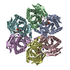
| Title | Crystal Structure of the Uridine Phosphorylase from Salmonella Typhimurium in Complex with Uracil and Phosphate Ion at 2.49A Resolution | ||||||
 Components Components | Uridine phosphorylase | ||||||
 Keywords Keywords | TRANSFERASE / uridine phosphorylase / Cytoplasm / Glycosyltransferase | ||||||
| Function / homology |  Function and homology information Function and homology informationnucleoside catabolic process / uridine phosphorylase / nucleotide catabolic process / UMP salvage / uridine phosphorylase activity / cytosolSimilarity search - Function | ||||||
| Biological species |  Salmonella typhimurium (bacteria) Salmonella typhimurium (bacteria) | ||||||
| Method | X-RAY DIFFRACTION / SYNCHROTRON / MOLECULAR REPLACEMENT / Resolution: 2.51 Å | ||||||
 Authors Authors | Timofeev, V.I. / Pavlyuk, B.P. / Lashkov, A.A. / Gabdoulkhakov, A.G. / Mikhailov, A.M. | ||||||
 Citation Citation | Journal: To be published Title: Crystal Structure of the Uridine Phosphorylase from Salmonella Typhimurium in Complex with Uracil and Phosphate Ion at 2.49A Resolution Authors: Timofeev, V.I. / Pavlyuk, B.P. / Lashkov, A.A. / Gabdoulkhakov, A.G. / Mikhailov, A.M. | ||||||
| History |
|
- Structure visualization
| Structure viewer | Molecule: MolmilJmol/JSmol |
|---|
- Downloads & links
-Download
| PDBx/mmCIF format | 2rj3.cif.gz | 287.1 KB | Display | PDBx/mmCIF format |
|---|---|---|---|---|
| PDB format | pdb2rj3.ent.gz | 230.4 KB | Display | PDB format |
| PDBx/mmJSON format | 2rj3.json.gz | Tree view | PDBx/mmJSON format | |
| Others |  Other downloads Other downloads |
-Validation report
| Arichive directory | https://data.pdbj.org/pub/pdb/validation_reports/rj/2rj3ftp://data.pdbj.org/pub/pdb/validation_reports/rj/2rj3 | HTTPS FTP |
|---|
-Related structure data
| Related structure data |  2oecS S: Starting model for refinement |
|---|---|
| Similar structure data |

-Links
 PDBj
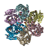
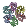
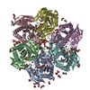
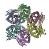
PDBj- Assembly
| Deposited unit | 
| ||||||||
|---|---|---|---|---|---|---|---|---|---|
| 1 |
| ||||||||
| Unit cell |
|
-Components
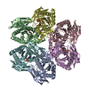
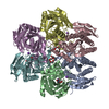
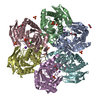
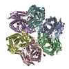
| #1: Protein | / UrdPase / UPase Mass: 27037.896 Da / Num. of mol.: 6 Source method: isolated from a genetically manipulated source Source: (gene. exp.) Salmonella typhimurium (bacteria) / Strain: LT2 / Gene: udp / Plasmid: PBLUESCRIPT IISK / Production host: Escherichia coli (E. coli) / Strain (production host): BL21, DE3 / References: UniProt: P0A1F6, uridine phosphorylase#2: Chemical | ChemComp-PO4 / Phosphate  Mass: 94.971 Da / Num. of mol.: 6 / Source method: obtained synthetically / Formula: PO4 Mass: 94.971 Da / Num. of mol.: 6 / Source method: obtained synthetically / Formula: PO4#3: Chemical | ChemComp-URA / Uracil  Mass: 112.087 Da / Num. of mol.: 5 / Source method: obtained synthetically / Formula: C4H4N2O2 Mass: 112.087 Da / Num. of mol.: 5 / Source method: obtained synthetically / Formula: C4H4N2O2#4: Water | ChemComp-HOH / | Water Mass: 18.015 Da / Num. of mol.: 218 / Source method: isolated from a natural source / Formula: H2O Mass: 18.015 Da / Num. of mol.: 218 / Source method: isolated from a natural source / Formula: H2O |
|---|
-Experimental details
-Experiment
| Experiment | Method: X-RAY DIFFRACTION / Number of used crystals: 1 |
|---|
- Sample preparation
| Crystal | Density Matthews: 2.28 Å3/Da / Density % sol: 46.12 % |
|---|---|
| Crystal grow | Temperature: 297 K / Method: vapor diffusion, hanging drop / Details: VAPOR DIFFUSION, HANGING DROP, temperature 297K |
-Data collection
| Diffraction | Mean temperature: 100 K |
|---|---|
| Diffraction source | Source: SYNCHROTRON / Site: EMBL/DESY, HAMBURG  / Beamline: X11 / Beamline: X11 |
| Detector | Type: MAR CCD 165 mm / Detector: CCD / Date: Sep 25, 2005 / Details: mirrors |
| Radiation | Protocol: SINGLE WAVELENGTH / Monochromatic (M) / Laue (L): M / Scattering type: x-ray |
| Radiation wavelength | Relative weight: 1 |
| Reflection | Resolution: 2.49→26.84 Å / Num. obs: 47321 |
- Processing
| Software | Name: REFMAC / Classification: refinement | ||||||||||||
|---|---|---|---|---|---|---|---|---|---|---|---|---|---|
| Refinement | Method to determine structure: MOLECULAR REPLACEMENT Starting model: PDB ENTRY 2OEC Resolution: 2.51→15.98 Å
| ||||||||||||
| Refinement step | Cycle: LAST / Resolution: 2.51→15.98 Å
|
