 Movie
Movie Controller

[English] 日本語
 Yorodumi
- PDB-2rj2: Crystal Structure of the Sugar Recognizing SCF Ubiquitin Ligase a... -
+ Open data

- Basic information
| Entry | Database: PDB / ID: 2rj2 | ||||||
|---|---|---|---|---|---|---|---|
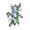
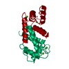
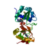
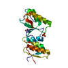
| Title | Crystal Structure of the Sugar Recognizing SCF Ubiquitin Ligase at 1.7 Resolution | ||||||
 Components Components | F-box only protein 2 | ||||||
 Keywords Keywords |  LIGASE / UBIQUITIN / SCF / UBIQUITIN LIGASE / LECTIN / Ubl conjugation pathway LIGASE / UBIQUITIN / SCF / UBIQUITIN LIGASE / LECTIN / Ubl conjugation pathway | ||||||
| Function / homology |  Function and homology informationextrinsic component of postsynaptic membrane / denatured protein binding / regulation of protein catabolic process at postsynapse, modulating synaptic transmission / glycoprotein catabolic process / Neddylation / Antigen processing: Ubiquitination & Proteasome degradation / SCF ubiquitin ligase complex / SCF-dependent proteasomal ubiquitin-dependent protein catabolic process / ERAD pathway / regulation of protein ubiquitination ...extrinsic component of postsynaptic membrane / denatured protein binding / regulation of protein catabolic process at postsynapse, modulating synaptic transmission / glycoprotein catabolic process / Neddylation / Antigen processing: Ubiquitination & Proteasome degradation / SCF ubiquitin ligase complex / SCF-dependent proteasomal ubiquitin-dependent protein catabolic process / ERAD pathway / regulation of protein ubiquitination / ubiquitin protein ligase activity / amyloid-beta binding / ubiquitin-dependent protein catabolic process / carbohydrate binding / dendritic spine / protein ubiquitination / negative regulation of cell population proliferation / glutamatergic synapse / endoplasmic reticulum / cytosol / cytoplasm Function and homology informationextrinsic component of postsynaptic membrane / denatured protein binding / regulation of protein catabolic process at postsynapse, modulating synaptic transmission / glycoprotein catabolic process / Neddylation / Antigen processing: Ubiquitination & Proteasome degradation / SCF ubiquitin ligase complex / SCF-dependent proteasomal ubiquitin-dependent protein catabolic process / ERAD pathway / regulation of protein ubiquitination ...extrinsic component of postsynaptic membrane / denatured protein binding / regulation of protein catabolic process at postsynapse, modulating synaptic transmission / glycoprotein catabolic process / Neddylation / Antigen processing: Ubiquitination & Proteasome degradation / SCF ubiquitin ligase complex / SCF-dependent proteasomal ubiquitin-dependent protein catabolic process / ERAD pathway / regulation of protein ubiquitination / ubiquitin protein ligase activity / amyloid-beta binding / ubiquitin-dependent protein catabolic process / carbohydrate binding / dendritic spine / protein ubiquitination / negative regulation of cell population proliferation / glutamatergic synapse / endoplasmic reticulum / cytosol / cytoplasmSimilarity search - Function | ||||||
| Biological species |  Mus musculus (house mouse) Mus musculus (house mouse) | ||||||
| Method | X-RAY DIFFRACTION / MOLECULAR REPLACEMENT / Resolution: 1.7 Å | ||||||
 Authors Authors | Vaijayanthimala, S. / Velmurugan, D. / Mizushima, T. / Yamane, T. / Yoshida, Y. / Tanaka, K. | ||||||
 Citation Citation | Journal: To be Published Title: Crystal Structure of the Sugar Recognizing SCF Ubiquitin Ligase at 1.7 Resolution Authors: Vaijayanthimala, S. / Velmurugan, D. / Mizushima, T. / Yamane, T. / Yoshida, Y. / Tanaka, K. #1: Journal: Nat.Struct.Mol.Biol. / Year: 2004Title: Structural basis of sugar-recognizing ubiquitin ligase Authors: Mizushima, T. / Hirao, T. / Yoshida, Y. / Lee, S.J. / Chiba, T. / Iwai, K. / Yamaguchi, Y. / Kato, K. / Tsukihara, T. / Tanaka, K. | ||||||
| History |
|
- Structure visualization
| Structure viewer | Molecule: MolmilJmol/JSmol |
|---|
- Downloads & links
-Download
| PDBx/mmCIF format | 2rj2.cif.gz | 58.3 KB | Display | PDBx/mmCIF format |
|---|---|---|---|---|
| PDB format | pdb2rj2.ent.gz | 40.7 KB | Display | PDB format |
| PDBx/mmJSON format | 2rj2.json.gz | Tree view | PDBx/mmJSON format | |
| Others |  Other downloads Other downloads |
-Validation report
| Arichive directory | https://data.pdbj.org/pub/pdb/validation_reports/rj/2rj2ftp://data.pdbj.org/pub/pdb/validation_reports/rj/2rj2 | HTTPS FTP |
|---|
-Related structure data
| Related structure data | 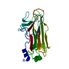 1umhS S: Starting model for refinement |
|---|---|
| Similar structure data |


-Links
 PDBj
PDBj


- Assembly
| Deposited unit | 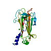
| ||||||||
|---|---|---|---|---|---|---|---|---|---|
| 1 |
| ||||||||
| Unit cell |
| ||||||||
| Components on special symmetry positions |
|
-Components
| #1: Protein | Mass: 21152.205 Da / Num. of mol.: 1 / Fragment: SBD DOMAIN. UNP residues 117-297 Source method: isolated from a genetically manipulated source Source: (gene. exp.) Mus musculus (house mouse) / Gene: Fbxo2, Fbx2 / Plasmid: PET15B / Production host:  Escherichia coli (E. coli) / Strain (production host): BL21(RIL) / References: UniProt: Q80UW2, ubiquitin-protein ligase Escherichia coli (E. coli) / Strain (production host): BL21(RIL) / References: UniProt: Q80UW2, ubiquitin-protein ligase |
|---|---|
| #2: Chemical | ChemComp-NI / Nickel  Mass: 58.693 Da / Num. of mol.: 1 / Source method: obtained synthetically / Formula: Ni Mass: 58.693 Da / Num. of mol.: 1 / Source method: obtained synthetically / Formula: Ni |
| #3: Chemical | ChemComp-CL / Chloride  Mass: 35.453 Da / Num. of mol.: 1 / Source method: obtained synthetically / Formula: Cl Mass: 35.453 Da / Num. of mol.: 1 / Source method: obtained synthetically / Formula: Cl |
| #4: Water | ChemComp-HOH / Water Mass: 18.015 Da / Num. of mol.: 256 / Source method: isolated from a natural source / Formula: H2O Mass: 18.015 Da / Num. of mol.: 256 / Source method: isolated from a natural source / Formula: H2O |
| Sequence details | THERE ARE DIFFERENCES BETWEEN THE REPORTED SEQRES AND THE DATABASE SEQUENCE. THE DEPOSITORS BELIEVE ...THERE ARE DIFFERENCE |
-Experimental details
-Experiment
| Experiment | Method: X-RAY DIFFRACTION / Number of used crystals: 1 |
|---|
- Sample preparation
| Crystal | Density Matthews: 3.04 Å3/Da / Density % sol: 59.6 % |
|---|---|
| Crystal grow | Temperature: 295 K / Method: vapor diffusion, hanging drop / pH: 8.5 Details: 0.1M TRIS, 0.1%(v/v) PEG 400, 0.01M NICKEL CHLORIDE, 1.7M AMMONIUM SULFATE, pH 8.5, VAPOR DIFFUSION, HANGING DROP, temperature 295K |
-Data collection
| Diffraction | Mean temperature: 100 K |
|---|---|
| Diffraction source | Source: ROTATING ANODE / Type: RIGAKU ULTRAX 18 / Wavelength: 1.5418 Å |
| Detector | Type: RIGAKU RAXIS IV / Detector: IMAGE PLATE / Date: Nov 23, 2006 |
| Radiation | Monochromator: Nickel filter / Protocol: SINGLE WAVELENGTH / Monochromatic (M) / Laue (L): M / Scattering type: x-ray |
| Radiation wavelength | Wavelength: 1.5418 Å / Relative weight: 1 |
| Reflection | Resolution: 1.7→100 Å / Num. all: 27682 / Num. obs: 27682 / % possible obs: 97.4 % / Rsym value: 0.072 |
| Reflection shell | Resolution: 1.7→1.79 Å / Rsym value: 0.241 / % possible all: 99.8 |
- Processing
| Software |
| |||||||||||||||||||||||||||||||||||||||||||||||||||||||||||||||||||||||||||||||||||||||||||||||
|---|---|---|---|---|---|---|---|---|---|---|---|---|---|---|---|---|---|---|---|---|---|---|---|---|---|---|---|---|---|---|---|---|---|---|---|---|---|---|---|---|---|---|---|---|---|---|---|---|---|---|---|---|---|---|---|---|---|---|---|---|---|---|---|---|---|---|---|---|---|---|---|---|---|---|---|---|---|---|---|---|---|---|---|---|---|---|---|---|---|---|---|---|---|---|---|---|
| Refinement | Method to determine structure: MOLECULAR REPLACEMENT Starting model: PDB ENTRY 1UMH Resolution: 1.7→19.91 Å / Cor.coef. Fo:Fc: 0.957 / Cor.coef. Fo:Fc free: 0.946 / SU B: 1.81 / SU ML: 0.061 / Cross valid method: THROUGHOUT / σ(F): 0 / ESU R: 0.105 / ESU R Free: 0.103 / Stereochemistry target values: MAXIMUM LIKELIHOOD
| |||||||||||||||||||||||||||||||||||||||||||||||||||||||||||||||||||||||||||||||||||||||||||||||
| Solvent computation | Ion probe radii: 0.8 Å / Shrinkage radii: 0.8 Å / VDW probe radii: 1.4 Å / Solvent model: MASK | |||||||||||||||||||||||||||||||||||||||||||||||||||||||||||||||||||||||||||||||||||||||||||||||
| Displacement parameters | Biso mean: 22.937 Å2
| |||||||||||||||||||||||||||||||||||||||||||||||||||||||||||||||||||||||||||||||||||||||||||||||
| Refinement step | Cycle: LAST / Resolution: 1.7→19.91 Å
| |||||||||||||||||||||||||||||||||||||||||||||||||||||||||||||||||||||||||||||||||||||||||||||||
| Refine LS restraints |
| |||||||||||||||||||||||||||||||||||||||||||||||||||||||||||||||||||||||||||||||||||||||||||||||
| LS refinement shell | Resolution: 1.7→1.744 Å / Total num. of bins used: 20
|
