 Movie
Movie Controller

[English] 日本語
 Yorodumi
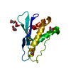
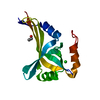
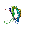
- PDB-2rea: Crystal structures of C2ALPHA-PI3 kinase PX-domain domain indicat... -
+ Open data

- Basic information
| Entry | Database: PDB / ID: 2rea | ||||||
|---|---|---|---|---|---|---|---|
| Title | Crystal structures of C2ALPHA-PI3 kinase PX-domain domain indicate conformational change associated with ligand binding. | ||||||
 Components Components | Phosphatidylinositol-4-phosphate 3-kinase C2 domain-containing alpha polypeptide | ||||||
 Keywords Keywords |  TRANSFERASE / PX DOMAIN / PI3K / KINASE / PHOSPHORYLATION / NUCLEAR PROTEIN / PHOSPHOINOSITIDE / Cytoplasm / Cytoplasmic vesicle / Golgi apparatus / Membrane / Nucleus / Polymorphism TRANSFERASE / PX DOMAIN / PI3K / KINASE / PHOSPHORYLATION / NUCLEAR PROTEIN / PHOSPHOINOSITIDE / Cytoplasm / Cytoplasmic vesicle / Golgi apparatus / Membrane / Nucleus / Polymorphism | ||||||
| Function / homology |  Function and homology information Function and homology informationvascular associated smooth muscle contraction / Synthesis of PIPs at the late endosome membrane / Synthesis of PIPs at the early endosome membrane / Synthesis of PIPs at the Golgi membrane / phosphatidylinositol-4-phosphate 3-kinase / clathrin coat assembly / membrane organization / phosphatidylinositol biosynthetic process / phosphatidylinositol 3-kinase complex / 1-phosphatidylinositol-4-phosphate 3-kinase activity ...vascular associated smooth muscle contraction / Synthesis of PIPs at the late endosome membrane / Synthesis of PIPs at the early endosome membrane / Synthesis of PIPs at the Golgi membrane / phosphatidylinositol-4-phosphate 3-kinase / clathrin coat assembly / membrane organization / phosphatidylinositol biosynthetic process / phosphatidylinositol 3-kinase complex / 1-phosphatidylinositol-4-phosphate 3-kinase activity / 1-phosphatidylinositol-4,5-bisphosphate 3-kinase activity / phosphatidylinositol-4,5-bisphosphate 3-kinase / phosphatidylinositol 3-kinase / clathrin-coated vesicle / phosphatidylinositol-3-phosphate biosynthetic process / 1-phosphatidylinositol-3-kinase activity / clathrin binding / positive regulation of cell migration involved in sprouting angiogenesis / Golgi Associated Vesicle Biogenesis / exocytosis / Synthesis of PIPs at the plasma membrane / platelet-derived growth factor receptor signaling pathway / positive regulation of autophagy / phosphatidylinositol binding / phosphatidylinositol 3-kinase/protein kinase B signal transduction / epidermal growth factor receptor signaling pathway / trans-Golgi network / endocytosis / insulin receptor signaling pathway / Clathrin-mediated endocytosis / vesicle / phosphorylation / intracellular membrane-bounded organelle / extracellular exosome / nucleoplasm / ATP binding / membrane / plasma membrane / cytosol / cytoplasmSimilarity search - Function | ||||||
| Biological species |  Homo sapiens (human) Homo sapiens (human) | ||||||
| Method | X-RAY DIFFRACTION / MOLECULAR REPLACEMENT / Resolution: 2.5 Å | ||||||
 Authors Authors | Parkinson, G.N. / Vines, D. / Driscoll, P.C. / Djordjevic, S. | ||||||
 Citation Citation | Journal: Bmc Struct.Biol. / Year: 2008 Title: Crystal structures of PI3K-C2alpha PX domain indicate conformational change associated with ligand binding Authors: Parkinson, G.N. / Vines, D. / Driscoll, P.C. / Djordjevic, S. | ||||||
| History |
|
- Structure visualization
| Structure viewer | Molecule: MolmilJmol/JSmol |
|---|
- Downloads & links
-Download
| PDBx/mmCIF format | 2rea.cif.gz | 37.7 KB | Display | PDBx/mmCIF format |
|---|---|---|---|---|
| PDB format | pdb2rea.ent.gz | 25.4 KB | Display | PDB format |
| PDBx/mmJSON format | 2rea.json.gz | Tree view | PDBx/mmJSON format | |
| Others |  Other downloads Other downloads |
-Validation report
| Arichive directory | https://data.pdbj.org/pub/pdb/validation_reports/re/2reaftp://data.pdbj.org/pub/pdb/validation_reports/re/2rea | HTTPS FTP |
|---|
-Related structure data
| Related structure data |  2redC 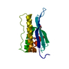 1ocsS C: citing same article ( S: Starting model for refinement |
|---|---|
| Similar structure data |


-Links
 PDBj
PDBj


- Assembly
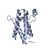
| Deposited unit | 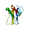
| ||||||||
|---|---|---|---|---|---|---|---|---|---|
| 1 |
| ||||||||
| Unit cell |
|
-Components
| #1: Protein | Mass: 14375.570 Da / Num. of mol.: 1 / Fragment: PX-DOMAIN, RESIDUES 1421-1532 Source method: isolated from a genetically manipulated source Source: (gene. exp.) Homo sapiens (human) / Gene: PIK3C2A / Plasmid: PET-21B / Production host:  Escherichia coli (E. coli) / Strain (production host): BL21(DE3)PLYSS Escherichia coli (E. coli) / Strain (production host): BL21(DE3)PLYSSReferences: UniProt: O00443, phosphatidylinositol-4-phosphate 3-kinase |
|---|---|
| #2: Water | ChemComp-HOH / Water Mass: 18.015 Da / Num. of mol.: 19 / Source method: isolated from a natural source / Formula: H2O Mass: 18.015 Da / Num. of mol.: 19 / Source method: isolated from a natural source / Formula: H2O |
-Experimental details
-Experiment
| Experiment | Method: X-RAY DIFFRACTION / Number of used crystals: 1 |
|---|
- Sample preparation
| Crystal | Density Matthews: 2.85 Å3/Da / Density % sol: 56.45 % |
|---|---|
| Crystal grow | Temperature: 293 K / Method: vapor diffusion, hanging drop / pH: 6 Details: 0.1M MALEIC ACID/NAOH, 10% GLYCEROL, PH 6.00, VAPOR DIFFUSION, HANGING DROP, TEMPERATURE 293K |
-Data collection
| Diffraction | Mean temperature: 105 K |
|---|---|
| Diffraction source | Source: ROTATING ANODE / Type: RIGAKU RU200 / Wavelength: 1.5418 / Wavelength: 1.5418 Å |
| Detector | Type: RIGAKU RAXIS IV / Detector: IMAGE PLATE / Date: Nov 4, 2004 / Details: MIRRORS OSMIC |
| Radiation | Protocol: SINGLE WAVELENGTH / Monochromatic (M) / Laue (L): M / Scattering type: x-ray |
| Radiation wavelength | Wavelength: 1.5418 Å / Relative weight: 1 |
| Reflection | Resolution: 2.5→30 Å / Num. all: 5731 / Num. obs: 5731 / % possible obs: 89.4 % / Observed criterion σ(F): 0 / Observed criterion σ(I): 0 / Redundancy: 3.5 % / Rmerge(I) obs: 0.028 / Net I/σ(I): 40 |
| Reflection shell | Resolution: 2.5→2.59 Å / Rmerge(I) obs: 0.107 / Mean I/σ(I) obs: 9 / % possible all: 70.9 |
- Processing
| Software |
| ||||||||||||||||||||||||||||||||||||||||||||||||||||||||||||||||||||||||||||||||||||||||||||||||||||||||||||||||||||||||||||||||||||||||||||||||||||||||||||||||||||||||||
|---|---|---|---|---|---|---|---|---|---|---|---|---|---|---|---|---|---|---|---|---|---|---|---|---|---|---|---|---|---|---|---|---|---|---|---|---|---|---|---|---|---|---|---|---|---|---|---|---|---|---|---|---|---|---|---|---|---|---|---|---|---|---|---|---|---|---|---|---|---|---|---|---|---|---|---|---|---|---|---|---|---|---|---|---|---|---|---|---|---|---|---|---|---|---|---|---|---|---|---|---|---|---|---|---|---|---|---|---|---|---|---|---|---|---|---|---|---|---|---|---|---|---|---|---|---|---|---|---|---|---|---|---|---|---|---|---|---|---|---|---|---|---|---|---|---|---|---|---|---|---|---|---|---|---|---|---|---|---|---|---|---|---|---|---|---|---|---|---|---|---|---|
| Refinement | Method to determine structure: MOLECULAR REPLACEMENT Starting model: PDB ENTRY 1OCS Resolution: 2.5→27.19 Å / Cor.coef. Fo:Fc: 0.918 / Cor.coef. Fo:Fc free: 0.84 / SU B: 8.757 / SU ML: 0.204 / Cross valid method: THROUGHOUT / σ(F): 0 / ESU R: 0.473 / ESU R Free: 0.352 / Stereochemistry target values: MAXIMUM LIKELIHOOD
| ||||||||||||||||||||||||||||||||||||||||||||||||||||||||||||||||||||||||||||||||||||||||||||||||||||||||||||||||||||||||||||||||||||||||||||||||||||||||||||||||||||||||||
| Solvent computation | Ion probe radii: 0.8 Å / Shrinkage radii: 0.8 Å / VDW probe radii: 1.4 Å / Solvent model: MASK | ||||||||||||||||||||||||||||||||||||||||||||||||||||||||||||||||||||||||||||||||||||||||||||||||||||||||||||||||||||||||||||||||||||||||||||||||||||||||||||||||||||||||||
| Displacement parameters | Biso mean: 40.838 Å2 | ||||||||||||||||||||||||||||||||||||||||||||||||||||||||||||||||||||||||||||||||||||||||||||||||||||||||||||||||||||||||||||||||||||||||||||||||||||||||||||||||||||||||||
| Refinement step | Cycle: LAST / Resolution: 2.5→27.19 Å
| ||||||||||||||||||||||||||||||||||||||||||||||||||||||||||||||||||||||||||||||||||||||||||||||||||||||||||||||||||||||||||||||||||||||||||||||||||||||||||||||||||||||||||
| Refine LS restraints |
| ||||||||||||||||||||||||||||||||||||||||||||||||||||||||||||||||||||||||||||||||||||||||||||||||||||||||||||||||||||||||||||||||||||||||||||||||||||||||||||||||||||||||||
| LS refinement shell | Resolution: 2.5→2.565 Å / Total num. of bins used: 20
|
