 Movie
Movie Controller

[English] 日本語
 Yorodumi
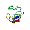
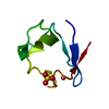
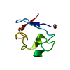
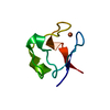
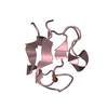
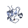
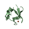
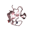
- PDB-2rdv: RUBREDOXIN FROM DESULFOVIBRIO VULGARIS MIYAZAKI F, MONOCLINIC CRY... -
+ Open data

- Basic information
| Entry | Database: PDB / ID: 2rdv | ||||||
|---|---|---|---|---|---|---|---|
| Title | RUBREDOXIN FROM DESULFOVIBRIO VULGARIS MIYAZAKI F, MONOCLINIC CRYSTAL FORM | ||||||
 Components Components | RUBREDOXIN | ||||||
 Keywords Keywords | ELECTRON TRANSFER / RUBREDOXIN / COMPND / ELECTRON TRANSFER PROTEIN / METALLOPROTEIN / SULFATE-REDUCING BACTERIUM | ||||||
| Function / homology |  Function and homology information Function and homology informationalkane catabolic process / electron transfer activity / iron ion binding / cytoplasmSimilarity search - Function | ||||||
| Biological species |  Desulfovibrio vulgaris str. 'Miyazaki F' (bacteria) Desulfovibrio vulgaris str. 'Miyazaki F' (bacteria) | ||||||
| Method | X-RAY DIFFRACTION / SYNCHROTRON / MOLECULAR REPLACEMENT / Resolution: 1.9 Å | ||||||
 Authors Authors | Higuchi, Y. / Yasuoka, N. | ||||||
 Citation Citation | Journal: Acta Crystallogr.,Sect.D / Year: 1999 Title: Structure determination of rubredoxin from Desulfovibrio vulgaris Miyazaki F in two crystal forms. Authors: Misaki, S. / Morimoto, Y. / Ogata, M. / Yagi, T. / Higuchi, Y. / Yasuoka, N. #1: Journal: Protein Pept.Lett. / Year: 1998Title: Preliminary Crystallographic Study of Two Crystal Forms of Rubredoxin from Sulfate-Reducing Bactrium Authors: Higuchi, Y. / Sugiyama, M.S. / Morimoto, S. / Ogata, Y. / Yagi, M. / Yasuoka, T. | ||||||
| History |
|
- Structure visualization
| Structure viewer | Molecule: MolmilJmol/JSmol |
|---|
- Downloads & links
-Download
| PDBx/mmCIF format | 2rdv.cif.gz | 40.6 KB | Display | PDBx/mmCIF format |
|---|---|---|---|---|
| PDB format | pdb2rdv.ent.gz | 28.9 KB | Display | PDB format |
| PDBx/mmJSON format | 2rdv.json.gz | Tree view | PDBx/mmJSON format | |
| Others |  Other downloads Other downloads |
-Validation report
| Arichive directory | https://data.pdbj.org/pub/pdb/validation_reports/rd/2rdvftp://data.pdbj.org/pub/pdb/validation_reports/rd/2rdv | HTTPS FTP |
|---|
-Related structure data
| Related structure data |  1rdvSC S: Starting model for refinement C: citing same article ( |
|---|---|
| Similar structure data |


-Links
 PDBj
PDBj
- Assembly
| Deposited unit | 
| ||||||||||||
|---|---|---|---|---|---|---|---|---|---|---|---|---|---|
| 1 | 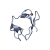
| ||||||||||||
| 2 | 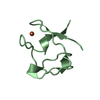
| ||||||||||||
| 3 | 
| ||||||||||||
| Unit cell |
| ||||||||||||
| Noncrystallographic symmetry (NCS) | NCS oper:
|
-Components
| #1: Protein | Mass: 5602.237 Da / Num. of mol.: 3 / Source method: isolated from a natural source / Details: IAM 12604 Source: (natural) Desulfovibrio vulgaris str. 'Miyazaki F' (bacteria)Species: Desulfovibrio vulgaris / Strain: MIYAZAKI F / References: UniProt: P15412#2: Chemical | Iron  Mass: 55.845 Da / Num. of mol.: 3 / Source method: obtained synthetically / Formula: Fe Mass: 55.845 Da / Num. of mol.: 3 / Source method: obtained synthetically / Formula: Fe#3: Water | ChemComp-HOH / | Water Mass: 18.015 Da / Num. of mol.: 86 / Source method: isolated from a natural source / Formula: H2O Mass: 18.015 Da / Num. of mol.: 86 / Source method: isolated from a natural source / Formula: H2O |
|---|
-Experimental details
-Experiment
| Experiment | Method: X-RAY DIFFRACTION / Number of used crystals: 1 |
|---|
- Sample preparation
| Crystal | Density Matthews: 1.87 Å3/Da / Density % sol: 34.2 % | |||||||||||||||||||||||||
|---|---|---|---|---|---|---|---|---|---|---|---|---|---|---|---|---|---|---|---|---|---|---|---|---|---|---|
| Crystal grow | pH: 7.4 / Details: pH 7.4 | |||||||||||||||||||||||||
| Crystal | *PLUS | |||||||||||||||||||||||||
| Crystal grow | *PLUS Temperature: 10 ℃ / pH: 7 / Method: microdialysis / Details: Higuchi, Y., (1998) Protein Pept.Lett., 5, 175. | |||||||||||||||||||||||||
| Components of the solutions | *PLUS
|
-Data collection
| Diffraction | Mean temperature: 288 K |
|---|---|
| Diffraction source | Source: SYNCHROTRON / Site: Photon Factory  / Beamline: BL-6A / Wavelength: 1 / Beamline: BL-6A / Wavelength: 1 |
| Detector | Type: FUJI / Detector: IMAGE PLATE / Details: MIRRORS |
| Radiation | Monochromator: SI(111) / Monochromatic (M) / Laue (L): M / Scattering type: x-ray |
| Radiation wavelength | Wavelength: 1 Å / Relative weight: 1 |
| Reflection | Resolution: 1.9→20 Å / Num. obs: 53316 / % possible obs: 82.5 % / Observed criterion σ(I): 0 / Rmerge(I) obs: 0.111 |
| Reflection | *PLUS Num. obs: 9604 / Observed criterion σ(I): 3 / Num. measured all: 53316 |
- Processing
| Software |
| ||||||||||||||||||||||||||||||||||||||||||||||||||||||||||||
|---|---|---|---|---|---|---|---|---|---|---|---|---|---|---|---|---|---|---|---|---|---|---|---|---|---|---|---|---|---|---|---|---|---|---|---|---|---|---|---|---|---|---|---|---|---|---|---|---|---|---|---|---|---|---|---|---|---|---|---|---|---|
| Refinement | Method to determine structure: MOLECULAR REPLACEMENT Starting model: PDB ENTRY 1RDV Resolution: 1.9→20 Å / Isotropic thermal model: RESTRAINED / σ(F): 3
| ||||||||||||||||||||||||||||||||||||||||||||||||||||||||||||
| Displacement parameters | Biso mean: 14.1 Å2 | ||||||||||||||||||||||||||||||||||||||||||||||||||||||||||||
| Refinement step | Cycle: LAST / Resolution: 1.9→20 Å
| ||||||||||||||||||||||||||||||||||||||||||||||||||||||||||||
| Refine LS restraints |
| ||||||||||||||||||||||||||||||||||||||||||||||||||||||||||||
| Software | *PLUS Name: X-PLOR / Classification: refinement | ||||||||||||||||||||||||||||||||||||||||||||||||||||||||||||
| Refine LS restraints | *PLUS
|
