 Movie
Movie Controller

[English] 日本語
 Yorodumi
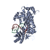
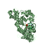
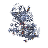
- PDB-2r8k: Structure of the Eukaryotic DNA Polymerase eta in complex with 1,... -
+ Open data

- Basic information
| Entry | Database: PDB / ID: 2r8k | ||||||
|---|---|---|---|---|---|---|---|
| Title | Structure of the Eukaryotic DNA Polymerase eta in complex with 1,2-d(GpG)-cisplatin containing DNA | ||||||
 Components Components |
| ||||||
 Keywords Keywords |  REPLICATION / TRANSFERASE/DNA / protein-cisplatin-DNA-dNTP complex / TRANSFERASE-DNA COMPLEX REPLICATION / TRANSFERASE/DNA / protein-cisplatin-DNA-dNTP complex / TRANSFERASE-DNA COMPLEX | ||||||
| Function / homology |  Function and homology information Function and homology informationmitotic sister chromatid cohesion / error-free translesion synthesis / error-prone translesion synthesis / replication fork / chromosome segregation / response to radiation / site of double-strand break / DNA replication / damaged DNA binding / DNA-directed DNA polymerase ...mitotic sister chromatid cohesion / error-free translesion synthesis / error-prone translesion synthesis / replication fork / chromosome segregation / response to radiation / site of double-strand break / DNA replication / damaged DNA binding / DNA-directed DNA polymerase / DNA-directed DNA polymerase activity / mitochondrion / metal ion binding / nucleusSimilarity search - Function | ||||||
| Biological species |  Saccharomyces cerevisiae (brewer's yeast) Saccharomyces cerevisiae (brewer's yeast) | ||||||
| Method | X-RAY DIFFRACTION / SYNCHROTRON / MOLECULAR REPLACEMENT / Resolution: 3.3 Å | ||||||
 Authors Authors | Carell, T. / Alt, A. / Lammens, K. | ||||||
 Citation Citation | Journal: Science / Year: 2007 Title: Bypass of DNA lesions generated during anticancer treatment with cisplatin by DNA polymerase eta Authors: Alt, A. / Lammens, K. / Chiocchini, C. / Lammens, A. / Pieck, J.C. / Kuch, D. / Hopfner, K.P. / Carell, T. | ||||||
| History |
|
- Structure visualization
| Structure viewer | Molecule: MolmilJmol/JSmol |
|---|
- Downloads & links
-Download
| PDBx/mmCIF format | 2r8k.cif.gz | 239.6 KB | Display | PDBx/mmCIF format |
|---|---|---|---|---|
| PDB format | pdb2r8k.ent.gz | 187 KB | Display | PDB format |
| PDBx/mmJSON format | 2r8k.json.gz | Tree view | PDBx/mmJSON format | |
| Others |  Other downloads Other downloads |
-Validation report
| Arichive directory | https://data.pdbj.org/pub/pdb/validation_reports/r8/2r8kftp://data.pdbj.org/pub/pdb/validation_reports/r8/2r8k | HTTPS FTP |
|---|
-Related structure data
| Related structure data |  2r8jC 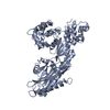 1jihS S: Starting model for refinement C: citing same article ( |
|---|---|
| Similar structure data |


-Links
 PDBj
PDBj


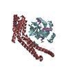
- Assembly
| Deposited unit | 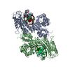
| ||||||||
|---|---|---|---|---|---|---|---|---|---|
| 1 | 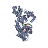
| ||||||||
| 2 | 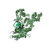
| ||||||||
| Unit cell |
|
-Components
-DNA chain , 2 types, 4 molecules QPUT
| #1: DNA chain | Mass: 2811.846 Da / Num. of mol.: 2 / Source method: obtained synthetically #2: DNA chain | Mass: 2989.970 Da / Num. of mol.: 2 / Source method: obtained synthetically |
|---|
-Protein , 1 types, 2 molecules AB
| #3: Protein | / E.C.2.7.7.7 / Radiation sensitive protein 30 Mass: 62617.781 Da / Num. of mol.: 2 / Fragment: Catalytic domain Source method: isolated from a genetically manipulated source Source: (gene. exp.) Saccharomyces cerevisiae (brewer's yeast)Strain: YPH499 (ATCC 76625) / Gene: RAD30, DBH1 / Plasmid: pExp007 / Production host:  Escherichia coli (E. coli) / Strain (production host): Bl21 DE3 Rosetta / References: UniProt: Q04049, DNA-directed DNA polymerase Escherichia coli (E. coli) / Strain (production host): Bl21 DE3 Rosetta / References: UniProt: Q04049, DNA-directed DNA polymerase |
|---|
-Non-polymers , 3 types, 8 molecules 


| #4: Chemical | Cisplatin Mass: 300.045 Da / Num. of mol.: 2 / Source method: obtained synthetically / Formula: Cl2H6N2Pt / Comment: medication, chemotherapy*YM Mass: 300.045 Da / Num. of mol.: 2 / Source method: obtained synthetically / Formula: Cl2H6N2Pt / Comment: medication, chemotherapy*YM#5: Chemical | ChemComp-CA /  Mass: 40.078 Da / Num. of mol.: 4 / Source method: obtained synthetically / Formula: Ca Mass: 40.078 Da / Num. of mol.: 4 / Source method: obtained synthetically / Formula: Ca#6: Chemical | Deoxyadenosine triphosphate Mass: 491.182 Da / Num. of mol.: 2 / Source method: obtained synthetically / Formula: C10H16N5O12P3 Mass: 491.182 Da / Num. of mol.: 2 / Source method: obtained synthetically / Formula: C10H16N5O12P3 |
|---|
-Experimental details
-Experiment
| Experiment | Method: X-RAY DIFFRACTION / Number of used crystals: 1 |
|---|
- Sample preparation
| Crystal | Density Matthews: 2.88 Å3/Da / Density % sol: 57.27 % | ||||||||||||||||||||
|---|---|---|---|---|---|---|---|---|---|---|---|---|---|---|---|---|---|---|---|---|---|
| Crystal grow | Temperature: 291 K / Method: vapor diffusion, hanging drop Details: 170 mM Calcium chloride, 16% PEG 3350, VAPOR DIFFUSION, HANGING DROP, temperature 291.0K | ||||||||||||||||||||
| Components of the solutions |
|
-Data collection
| Diffraction | Mean temperature: 100 K |
|---|---|
| Diffraction source | Source: SYNCHROTRON / Site: ESRF  / Beamline: ID23-1 / Wavelength: 1.0719 Å / Beamline: ID23-1 / Wavelength: 1.0719 Å |
| Detector | Type: ADSC QUANTUM 315 / Detector: CCD / Date: Feb 5, 2007 |
| Radiation | Monochromator: Si / Protocol: SINGLE WAVELENGTH / Monochromatic (M) / Laue (L): M / Scattering type: x-ray |
| Radiation wavelength | Wavelength: 1.0719 Å / Relative weight: 1 |
| Reflection | Resolution: 3.3→25 Å / Num. all: 24858 / Num. obs: 24858 / % possible obs: 98.4 % / Observed criterion σ(I): -3 / Biso Wilson estimate: 37.602 Å2 / Rsym value: 0.147 / Net I/σ(I): 11.78 |
- Processing
| Software |
| ||||||||||||||||||||||||||||
|---|---|---|---|---|---|---|---|---|---|---|---|---|---|---|---|---|---|---|---|---|---|---|---|---|---|---|---|---|---|
| Refinement | Method to determine structure: MOLECULAR REPLACEMENT Starting model: 1JIH Resolution: 3.3→25 Å / Isotropic thermal model: anisotropic / Cross valid method: THROUGHOUT / σ(F): 0 / σ(I): 0 / Stereochemistry target values: Engh & Huber
| ||||||||||||||||||||||||||||
| Displacement parameters | Biso mean: 35.59 Å2 | ||||||||||||||||||||||||||||
| Refinement step | Cycle: LAST / Resolution: 3.3→25 Å
| ||||||||||||||||||||||||||||
| Refine LS restraints |
|
