 Movie
Movie Controller

[English] 日本語
 Yorodumi
- PDB-2r4l: Crystal structure of the long-chain fatty acid transporter FadL m... -
+ Open data

- Basic information
| Entry | Database: PDB / ID: 2r4l | ||||||
|---|---|---|---|---|---|---|---|
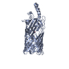
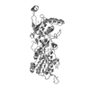
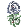
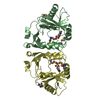
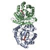
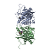
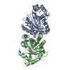
| Title | Crystal structure of the long-chain fatty acid transporter FadL mutant P34A | ||||||
 Components Components | Long-chain fatty acid transport protein | ||||||
 Keywords Keywords |  TRANSPORT PROTEIN / beta barrel / outer membrane protein / Lipid transport / Phage recognition / Transmembrane / Transport TRANSPORT PROTEIN / beta barrel / outer membrane protein / Lipid transport / Phage recognition / Transmembrane / Transport | ||||||
| Function / homology |  Function and homology information Function and homology informationlong-chain fatty acid transporting porin activity / ligand-gated channel activity / cell outer membraneSimilarity search - Function | ||||||
| Biological species |  Escherichia coli (E. coli) Escherichia coli (E. coli) | ||||||
| Method | X-RAY DIFFRACTION / SYNCHROTRON / MOLECULAR REPLACEMENT / Resolution: 3.3 Å | ||||||
 Authors Authors | Hearn, E.M. / Patel, D.R. / Lepore, B.W. / Indic, M. / van den Berg, B. | ||||||
 Citation Citation | Journal: Nature / Year: 2009 Title: Transmembrane passage of hydrophobic compounds through a protein channel wall Authors: Hearn, E.M. / Patel, D.R. / Lepore, B.W. / Indic, M. / van den Berg, B. #1: Journal: Science / Year: 2004Title: Crystal structure of the long-chain fatty acid transporter FadL Authors: van den Berg, B. / Black, P.N. / Clemons Jr., W.M. / Rapoport, T.A. | ||||||
| History |
|
- Structure visualization
| Structure viewer | Molecule: MolmilJmol/JSmol |
|---|
- Downloads & links
-Download
| PDBx/mmCIF format | 2r4l.cif.gz | 244 KB | Display | PDBx/mmCIF format |
|---|---|---|---|---|
| PDB format | pdb2r4l.ent.gz | 197.4 KB | Display | PDB format |
| PDBx/mmJSON format | 2r4l.json.gz | Tree view | PDBx/mmJSON format | |
| Others |  Other downloads Other downloads |
-Validation report
| Arichive directory | https://data.pdbj.org/pub/pdb/validation_reports/r4/2r4lftp://data.pdbj.org/pub/pdb/validation_reports/r4/2r4l | HTTPS FTP |
|---|
-Related structure data
| Related structure data | 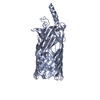 2r4nC 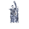 2r4oC  2r4pC  2r88C  3dwnC  3dwoC  1t16S  2r4m S: Starting model for refinement C: citing same article ( |
|---|---|
| Similar structure data |


-Links
 PDBj
PDBj- Assembly
| Deposited unit | 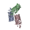
| ||||||||
|---|---|---|---|---|---|---|---|---|---|
| 1 | 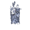
| ||||||||
| 2 | 
| ||||||||
| 3 | 
| ||||||||
| Unit cell |
|
-Components
| #1: Protein | Mass: 46728.902 Da / Num. of mol.: 3 / Mutation: P34A,I197T Source method: isolated from a genetically manipulated source Source: (gene. exp.) Escherichia coli (E. coli) / Gene: fadL, ttr / Plasmid: pBAD22 / Production host: Escherichia coli (E. coli) / Strain (production host): C43(DE3) / References: UniProt: P10384#2: Chemical | ChemComp-LDA / Lauryldimethylamine oxide  Mass: 229.402 Da / Num. of mol.: 6 / Source method: obtained synthetically / Formula: C14H31NO / Comment: LDAO, detergent*YM Mass: 229.402 Da / Num. of mol.: 6 / Source method: obtained synthetically / Formula: C14H31NO / Comment: LDAO, detergent*YM |
|---|
-Experimental details
-Experiment
| Experiment | Method: X-RAY DIFFRACTION / Number of used crystals: 1 |
|---|
- Sample preparation
| Crystal | Density Matthews: 3.38 Å3/Da / Density % sol: 63.56 % |
|---|---|
| Crystal grow | Temperature: 295 K / pH: 4.6 Details: 30% PEG 4000, 0.2M ammonium acetate, 0.1M sodium acetate, pH 4.60, VAPOR DIFFUSION, HANGING DROP, temperature 295K |
-Data collection
| Diffraction | Mean temperature: 100 K |
|---|---|
| Diffraction source | Source: SYNCHROTRON / Site: NSLS  / Beamline: X6A / Wavelength: 1.1 / Beamline: X6A / Wavelength: 1.1 |
| Detector | Type: ADSC QUANTUM 210 / Detector: CCD / Date: Jun 10, 2005 |
| Radiation | Monochromator: SI(111) CHANNEL CUT MONOCHROMATOR / Protocol: SINGLE WAVELENGTH / Monochromatic (M) / Laue (L): M / Scattering type: x-ray |
| Radiation wavelength | Wavelength: 1.1 Å / Relative weight: 1 |
| Reflection | Resolution: 3.3→48.4 Å / Num. obs: 27032 / % possible obs: 94.8 % / Observed criterion σ(I): 2 / Redundancy: 5.2 % / Rmerge(I) obs: 0.163 / Net I/σ(I): 5.1 |
| Reflection shell | Resolution: 3.3→3.42 Å / Redundancy: 5.1 % / Rmerge(I) obs: 0.501 / Mean I/σ(I) obs: 2.7 / Rsym value: 0.501 / % possible all: 95.9 |
- Processing
| Software |
| ||||||||||||||||||||||||||||||||||||||||||||||||||||||||||||
|---|---|---|---|---|---|---|---|---|---|---|---|---|---|---|---|---|---|---|---|---|---|---|---|---|---|---|---|---|---|---|---|---|---|---|---|---|---|---|---|---|---|---|---|---|---|---|---|---|---|---|---|---|---|---|---|---|---|---|---|---|---|
| Refinement | Method to determine structure: MOLECULAR REPLACEMENT Starting model: PDB ENTRY 1T16 Resolution: 3.3→10 Å / Cross valid method: THROUGHOUT / σ(F): 0 / Stereochemistry target values: ENGH & HUBER
| ||||||||||||||||||||||||||||||||||||||||||||||||||||||||||||
| Displacement parameters | Biso mean: 23.75 Å2
| ||||||||||||||||||||||||||||||||||||||||||||||||||||||||||||
| Refine analyze |
| ||||||||||||||||||||||||||||||||||||||||||||||||||||||||||||
| Refinement step | Cycle: LAST / Resolution: 3.3→10 Å
| ||||||||||||||||||||||||||||||||||||||||||||||||||||||||||||
| Refine LS restraints |
| ||||||||||||||||||||||||||||||||||||||||||||||||||||||||||||
| LS refinement shell | Resolution: 3.3→3.44 Å / Rfactor Rfree error: 0.019
|
