 Movie
Movie Controller

[English] 日本語
 Yorodumi
- PDB-2r3y: Crystal structure of the DegS protease in complex with the YWF ac... -
+ Open data

- Basic information
| Entry | Database: PDB / ID: 2r3y | ||||||
|---|---|---|---|---|---|---|---|
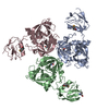
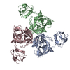
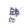
| Title | Crystal structure of the DegS protease in complex with the YWF activating peptide | ||||||
 Components Components |
| ||||||
 Keywords Keywords | hydrolase/hydrolase activator / reversible activation of a protease /  catalytic triad / Hydrolase / Periplasm / Serine protease / hydrolase-hydrolase activator COMPLEX catalytic triad / Hydrolase / Periplasm / Serine protease / hydrolase-hydrolase activator COMPLEX | ||||||
| Function / homology |  Function and homology informationpeptidase Do / cellular response to misfolded protein / serine-type peptidase activity / outer membrane-bounded periplasmic space / peptidase activity / serine-type endopeptidase activity / proteolysis / identical protein binding / plasma membrane Function and homology informationpeptidase Do / cellular response to misfolded protein / serine-type peptidase activity / outer membrane-bounded periplasmic space / peptidase activity / serine-type endopeptidase activity / proteolysis / identical protein binding / plasma membraneSimilarity search - Function | ||||||
| Biological species |  Escherichia coli (E. coli) Escherichia coli (E. coli) | ||||||
| Method | X-RAY DIFFRACTION / MOLECULAR REPLACEMENT / Resolution: 2.5 Å | ||||||
 Authors Authors | Clausen, T. / Hasselblatt, H. | ||||||
 Citation Citation | Journal: Genes Dev. / Year: 2007 Title: Regulation of the sigmaE stress response by DegS: how the PDZ domain keeps the protease inactive in the resting state and allows integration of different OMP-derived stress signals upon folding stress. Authors: Hasselblatt, H. / Kurzbauer, R. / Wilken, C. / Krojer, T. / Sawa, J. / Kurt, J. / Kirk, R. / Hasenbein, S. / Ehrmann, M. / Clausen, T. | ||||||
| History |
|
- Structure visualization
| Structure viewer | Molecule: MolmilJmol/JSmol |
|---|
- Downloads & links
-Download
| PDBx/mmCIF format | 2r3y.cif.gz | 169.4 KB | Display | PDBx/mmCIF format |
|---|---|---|---|---|
| PDB format | pdb2r3y.ent.gz | 134.4 KB | Display | PDB format |
| PDBx/mmJSON format | 2r3y.json.gz | Tree view | PDBx/mmJSON format | |
| Others |  Other downloads Other downloads |
-Validation report
| Arichive directory | https://data.pdbj.org/pub/pdb/validation_reports/r3/2r3yftp://data.pdbj.org/pub/pdb/validation_reports/r3/2r3y | HTTPS FTP |
|---|
-Related structure data
| Related structure data | 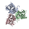 2r3uC 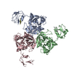 1sozS S: Starting model for refinement C: citing same article ( |
|---|---|
| Similar structure data |


-Links
 PDBj
PDBj
- Assembly
| Deposited unit | 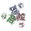
| ||||||||
|---|---|---|---|---|---|---|---|---|---|
| 1 |
| ||||||||
| Unit cell |
| ||||||||
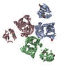
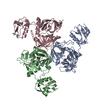
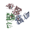
| Details | One homotrimer of DegS was observed in the asymmetric unit and should represent its physiological state. |
-Components
| #1: Protein | Mass: 33243.664 Da / Num. of mol.: 3 Source method: isolated from a genetically manipulated source Source: (gene. exp.) Escherichia coli (E. coli) / Gene: degS, hhoB, htrH / Plasmid: pET15-b / Species (production host): Escherichia coli / Production host: Escherichia coli BL21(DE3) (bacteria) / Strain (production host): BL21(DE3)References: UniProt: P0AEE3, Hydrolases; Acting on peptide bonds (peptidases); Serine endopeptidases#2: Protein/peptide | Mass: 1283.455 Da / Num. of mol.: 3 / Source method: obtained synthetically Details: The YWF peptide mimics the C-terminus of Outer Membrane Proteins. #3: Water | ChemComp-HOH / | Water Mass: 18.015 Da / Num. of mol.: 157 / Source method: isolated from a natural source / Formula: H2O Mass: 18.015 Da / Num. of mol.: 157 / Source method: isolated from a natural source / Formula: H2O |
|---|
-Experimental details
-Experiment
| Experiment | Method: X-RAY DIFFRACTION / Number of used crystals: 1 |
|---|
- Sample preparation
| Crystal | Density Matthews: 2.91 Å3/Da / Density % sol: 57.77 % |
|---|---|
| Crystal grow | Temperature: 292 K / Method: vapor diffusion, sitting drop / pH: 7.5 Details: The YWF (100 microM) was added to DegS and incubated 30 minutes before setting up the co-crystallization trials. Crystals of the complex were grown in sitting drops at 19 C by mixing 4 ...Details: The YWF (100 microM) was added to DegS and incubated 30 minutes before setting up the co-crystallization trials. Crystals of the complex were grown in sitting drops at 19 C by mixing 4 microL of DegS/YWF with 2 microL of a crystallization solution containing 0.1 M HEPES (pH 7.5), 6% PEG 6000, 9% MPD and 10 mM MgCl2. Crystal trials were set up in cryschem plates with a reservoir volume of 400 microL. , VAPOR DIFFUSION, SITTING DROP, temperature 292K |
-Data collection
| Diffraction | Mean temperature: 100 K |
|---|---|
| Diffraction source | Source: ROTATING ANODE / Type: RIGAKU RU300 / Wavelength: 1.5418 Å |
| Detector | Type: MAR scanner 345 mm plate / Detector: IMAGE PLATE / Date: Oct 30, 2006 / Details: mirrors |
| Radiation | Monochromator: Ni filter / Protocol: SINGLE WAVELENGTH / Monochromatic (M) / Laue (L): M / Scattering type: x-ray |
| Radiation wavelength | Wavelength: 1.5418 Å / Relative weight: 1 |
| Reflection | Resolution: 2.5→30 Å / Num. all: 39228 / Num. obs: 39228 / % possible obs: 95 % / Observed criterion σ(F): 0 / Observed criterion σ(I): 0 / Redundancy: 2.8 % / Biso Wilson estimate: 60 Å2 / Rmerge(I) obs: 0.044 / Rsym value: 0.047 / Net I/σ(I): 13.2 |
| Reflection shell | Resolution: 2.5→2.59 Å / Redundancy: 2.6 % / Rmerge(I) obs: 0.32 / Mean I/σ(I) obs: 2 / Num. unique all: 3644 / Rsym value: 0.33 / % possible all: 88.2 |
- Processing
| Software |
| |||||||||||||||||||||||||
|---|---|---|---|---|---|---|---|---|---|---|---|---|---|---|---|---|---|---|---|---|---|---|---|---|---|---|
| Refinement | Method to determine structure: MOLECULAR REPLACEMENT Starting model: PDB entry 1soz Resolution: 2.5→15 Å / Cross valid method: THROUGHOUT / σ(F): 0 / σ(I): 0 / Stereochemistry target values: Engh & Huber
| |||||||||||||||||||||||||
| Refinement step | Cycle: LAST / Resolution: 2.5→15 Å
| |||||||||||||||||||||||||
| Refine LS restraints |
|
