 Movie
Movie Controller

[English] 日本語
 Yorodumi
- PDB-2qvc: Crystal structure of a periplasmic sugar ABC transporter from The... -
+ Open data

- Basic information
| Entry | Database: PDB / ID: 2qvc | ||||||
|---|---|---|---|---|---|---|---|
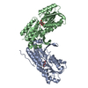
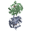
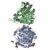
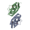
| Title | Crystal structure of a periplasmic sugar ABC transporter from Thermotoga maritima | ||||||
 Components Components | Sugar ABC transporter, periplasmic sugar-binding protein | ||||||
 Keywords Keywords | TRANSPORT PROTEIN / ABC sugar transporter / Sugar-binding protein / Protein Structure Initiative II / PSI II / NYSGXRC / 11013q / Structural Genomics / New York SGX Research Center for Structural Genomics | ||||||
| Function / homology |  Function and homology information Function and homology information | ||||||
| Biological species |   Thermotoga maritima MSB8 (bacteria) Thermotoga maritima MSB8 (bacteria) | ||||||
| Method |  X-RAY DIFFRACTION / SYNCHROTRON / SAD / Resolution: 2.4 Å X-RAY DIFFRACTION / SYNCHROTRON / SAD / Resolution: 2.4 Å | ||||||
 Authors Authors | Palani, K. / Kumaran, D. / Burley, S.K. / Swaminathan, S. / New York SGX Research Center for Structural Genomics (NYSGXRC) | ||||||
 Citation Citation | Journal: Acta Crystallogr.,Sect.F / Year: 2012 Title: Structure of a periplasmic glucose-binding protein from Thermotoga maritima. Authors: Palani, K. / Kumaran, D. / Burley, S.K. / Swaminathan, S. | ||||||
| History |
|
- Structure visualization
| Structure viewer | Molecule: MolmilJmol/JSmol |
|---|
- Downloads & links
-Download
| PDBx/mmCIF format | 2qvc.cif.gz | 234.2 KB | Display | PDBx/mmCIF format |
|---|---|---|---|---|
| PDB format | pdb2qvc.ent.gz | 198.8 KB | Display | PDB format |
| PDBx/mmJSON format | 2qvc.json.gz | Tree view | PDBx/mmJSON format | |
| Others |  Other downloads Other downloads |
-Validation report
| Summary document | 2qvc_validation.pdf.gz | 471.9 KB | Display | wwPDB validaton report |
|---|---|---|---|---|
| Full document | 2qvc_full_validation.pdf.gz | 490 KB | Display | |
| Data in XML | 2qvc_validation.xml.gz | 45.7 KB | Display | |
| Data in CIF | 2qvc_validation.cif.gz | 63.7 KB | Display | |
| Arichive directory | https://data.pdbj.org/pub/pdb/validation_reports/qv/2qvcftp://data.pdbj.org/pub/pdb/validation_reports/qv/2qvc | HTTPS FTP |
-Related structure data
| Similar structure data | |
|---|---|
| Other databases |


-Links
 PDBj
PDBj


- Assembly
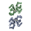
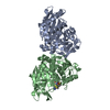
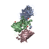
| Deposited unit | 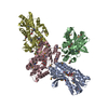
| ||||||||
|---|---|---|---|---|---|---|---|---|---|
| 1 | 
| ||||||||
| 2 | 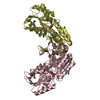
| ||||||||
| Unit cell |
|
-Components
| #1: Protein | Mass: 34447.680 Da / Num. of mol.: 4 / Fragment: Residues 33-334 Source method: isolated from a genetically manipulated source Source: (gene. exp.) Thermotoga maritima MSB8 (bacteria) / Species: Thermotoga maritima / Strain: MSB8, DSM 3109, JCM 10099 / Gene: TM_0114 / Plasmid: pSGX3(BC) / Species (production host): Escherichia coli / Production host: #2: Sugar | ChemComp-BGC /   Type: D-saccharide, beta linking / Mass: 180.156 Da / Num. of mol.: 4 Type: D-saccharide, beta linking / Mass: 180.156 Da / Num. of mol.: 4Source method: isolated from a genetically manipulated source Formula: C6H12O6 #3: Water | ChemComp-HOH / |  Mass: 18.015 Da / Num. of mol.: 287 / Source method: isolated from a natural source / Formula: H2O Mass: 18.015 Da / Num. of mol.: 287 / Source method: isolated from a natural source / Formula: H2O |
|---|
-Experimental details
-Experiment
| Experiment | Method: X-RAY DIFFRACTION / Number of used crystals: 1 |
|---|
- Sample preparation
| Crystal | Density Matthews: 2.98 Å3/Da / Density % sol: 58.73 % |
|---|---|
| Crystal grow | Temperature: 298 K / Method: vapor diffusion, sitting drop Details: 30% PEG 4000, 0.2 M Ammonium sulfate, VAPOR DIFFUSION, SITTING DROP, temperature 298.0K |
-Data collection
| Diffraction | Mean temperature: 100 K |
|---|---|
| Diffraction source | Source: SYNCHROTRON / Site: NSLS  / Beamline: X29A / Wavelength: 0.979 Å / Beamline: X29A / Wavelength: 0.979 Å |
| Detector | Type: ADSC QUANTUM 315 / Detector: CCD / Date: Jul 28, 2007 / Details: mirrors |
| Radiation | Monochromator: Si (111) / Protocol: SINGLE WAVELENGTH / Monochromatic (M) / Laue (L): M / Scattering type: x-ray |
| Radiation wavelength | Wavelength: 0.979 Å / Relative weight: 1 |
| Reflection | Resolution: 2.4→50 Å / Num. all: 60160 / Num. obs: 60160 / % possible obs: 96 % / Observed criterion σ(F): 0 / Observed criterion σ(I): 0 / Redundancy: 9.7 % / Biso Wilson estimate: 28.5 Å2 / Rmerge(I) obs: 0.096 / Net I/σ(I): 13 |
| Reflection shell | Resolution: 2.4→2.49 Å / Redundancy: 3.4 % / Rmerge(I) obs: 0.39 / Mean I/σ(I) obs: 2.5 / Num. unique all: 4512 / % possible all: 72.3 |
- Processing
| Software |
| ||||||||||||||||||||||||||||||||||||||||||||||||||||||||||||
|---|---|---|---|---|---|---|---|---|---|---|---|---|---|---|---|---|---|---|---|---|---|---|---|---|---|---|---|---|---|---|---|---|---|---|---|---|---|---|---|---|---|---|---|---|---|---|---|---|---|---|---|---|---|---|---|---|---|---|---|---|---|
| Refinement | Method to determine structure: SAD / Resolution: 2.4→30.83 Å / Rfactor Rfree error: 0.004 / Data cutoff high absF: 48519.07 / Data cutoff low absF: 0 / Isotropic thermal model: RESTRAINED / Cross valid method: THROUGHOUT / σ(F): 0 / Stereochemistry target values: Engh & Huber Details: Residues listed as missing in Remark 465 are due to lack of electron density. Residues with missing atoms listed in Remark 470 are due to lack of electron density for side chains and modeled as alanines.
| ||||||||||||||||||||||||||||||||||||||||||||||||||||||||||||
| Solvent computation | Solvent model: FLAT MODEL / Bsol: 24.7416 Å2 / ksol: 0.320539 e/Å3 | ||||||||||||||||||||||||||||||||||||||||||||||||||||||||||||
| Displacement parameters | Biso mean: 33.2 Å2
| ||||||||||||||||||||||||||||||||||||||||||||||||||||||||||||
| Refine analyze |
| ||||||||||||||||||||||||||||||||||||||||||||||||||||||||||||
| Refinement step | Cycle: LAST / Resolution: 2.4→30.83 Å
| ||||||||||||||||||||||||||||||||||||||||||||||||||||||||||||
| Refine LS restraints |
| ||||||||||||||||||||||||||||||||||||||||||||||||||||||||||||
| LS refinement shell | Resolution: 2.4→2.55 Å / Rfactor Rfree error: 0.016 / Total num. of bins used: 6
| ||||||||||||||||||||||||||||||||||||||||||||||||||||||||||||
| Xplor file |
|
