 Movie
Movie Controller

[English] 日本語
 Yorodumi
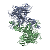
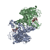
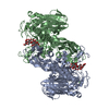
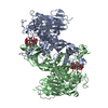
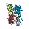
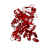
- PDB-2quq: Crystal Structure of the Essential Inner Kinetochore Protein Cep3p -
+ Open data

- Basic information
| Entry | Database: PDB / ID: 2quq | ||||||
|---|---|---|---|---|---|---|---|
| Title | Crystal Structure of the Essential Inner Kinetochore Protein Cep3p | ||||||
 Components Components | Centromere DNA-binding protein complex CBF3 subunit B | ||||||
 Keywords Keywords |  DNA BINDING PROTEIN / STRUCTURAL PROTEIN / dimer / Centromere / Chromosomal protein / DNA-binding / Metal-binding / Nucleus / Phosphorylation / Zinc / PROTEIN BINDING / CELL CYCLE DNA BINDING PROTEIN / STRUCTURAL PROTEIN / dimer / Centromere / Chromosomal protein / DNA-binding / Metal-binding / Nucleus / Phosphorylation / Zinc / PROTEIN BINDING / CELL CYCLE | ||||||
| Function / homology |  Function and homology information Function and homology informationCBF3 complex / septin ring assembly / centromeric DNA binding / kinetochore assembly / DNA binding, bending / mitotic spindle assembly checkpoint signaling / kinetochore / DNA-binding transcription factor activity, RNA polymerase II-specific / zinc ion binding / identical protein binding / nucleusSimilarity search - Function | ||||||
| Biological species |  Saccharomyces cerevisiae (brewer's yeast) Saccharomyces cerevisiae (brewer's yeast) | ||||||
| Method | X-RAY DIFFRACTION / SYNCHROTRON / MIRAS / Resolution: 2.8 Å | ||||||
 Authors Authors | Bellizzi III, J.J. / Harrison, S.C. | ||||||
 Citation Citation | Journal: Structure / Year: 2007 Title: Crystal structure of the yeast inner kinetochore subunit Cep3p. Authors: Bellizzi, J.J. / Sorger, P.K. / Harrison, S.C. | ||||||
| History |
|
- Structure visualization
| Structure viewer | Molecule: MolmilJmol/JSmol |
|---|
- Downloads & links
-Download
| PDBx/mmCIF format | 2quq.cif.gz | 120 KB | Display | PDBx/mmCIF format |
|---|---|---|---|---|
| PDB format | pdb2quq.ent.gz | 92.4 KB | Display | PDB format |
| PDBx/mmJSON format | 2quq.json.gz | Tree view | PDBx/mmJSON format | |
| Others |  Other downloads Other downloads |
-Validation report
| Arichive directory | https://data.pdbj.org/pub/pdb/validation_reports/qu/2quqftp://data.pdbj.org/pub/pdb/validation_reports/qu/2quq | HTTPS FTP |
|---|
-Related structure data
| Similar structure data |
|---|


-Links
 PDBj
PDBj- Assembly
| Deposited unit | 
| ||||||||
|---|---|---|---|---|---|---|---|---|---|
| 1 | 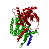
| ||||||||
| Unit cell |
| ||||||||
| Details | The biological unit is a dimer generated from the asymmetric unit by the crystallographic dyad. |
-Components
| #1: Protein | Mass: 66094.523 Da / Num. of mol.: 1 / Fragment: residues 47-608 Source method: isolated from a genetically manipulated source Source: (gene. exp.) Saccharomyces cerevisiae (brewer's yeast)Gene: CBF3B, CEP3 / Plasmid: pET3aTr / Production host:  Escherichia coli (E. coli) / Strain (production host): Rosetta(DE3)pLysS / References: UniProt: P40969 Escherichia coli (E. coli) / Strain (production host): Rosetta(DE3)pLysS / References: UniProt: P40969 |
|---|---|
| #2: Water | ChemComp-HOH / Water Mass: 18.015 Da / Num. of mol.: 15 / Source method: isolated from a natural source / Formula: H2O Mass: 18.015 Da / Num. of mol.: 15 / Source method: isolated from a natural source / Formula: H2O |
-Experimental details
-Experiment
| Experiment | Method: X-RAY DIFFRACTION / Number of used crystals: 3 |
|---|
- Sample preparation
| Crystal | Density Matthews: 3.13 Å3/Da / Density % sol: 60.68 % |
|---|---|
| Crystal grow | Temperature: 298 K / Method: vapor diffusion, hanging drop / pH: 7.5 Details: 100 mM HEPES, 5% PEG 4000, 500 mM NaCl, pH 7.5, vapor diffusion, hanging drop, temperature 298K |
-Data collection
| Diffraction |
| |||||||||||||||||||||||||||||||||||||||||||||||||||||||||||||||||||||||||||||
|---|---|---|---|---|---|---|---|---|---|---|---|---|---|---|---|---|---|---|---|---|---|---|---|---|---|---|---|---|---|---|---|---|---|---|---|---|---|---|---|---|---|---|---|---|---|---|---|---|---|---|---|---|---|---|---|---|---|---|---|---|---|---|---|---|---|---|---|---|---|---|---|---|---|---|---|---|---|---|
| Diffraction source |
| |||||||||||||||||||||||||||||||||||||||||||||||||||||||||||||||||||||||||||||
| Detector |
| |||||||||||||||||||||||||||||||||||||||||||||||||||||||||||||||||||||||||||||
| Radiation |
| |||||||||||||||||||||||||||||||||||||||||||||||||||||||||||||||||||||||||||||
| Radiation wavelength |
| |||||||||||||||||||||||||||||||||||||||||||||||||||||||||||||||||||||||||||||
| Reflection | Redundancy: 6.8 % / Av σ(I) over netI: 9.8 / Number: 139991 / Rmerge(I) obs: 0.072 / Χ2: 1.09 / D res high: 2.8 Å / D res low: 50 Å / Num. obs: 20443 / % possible obs: 94.2 | |||||||||||||||||||||||||||||||||||||||||||||||||||||||||||||||||||||||||||||
| Diffraction reflection shell |
| |||||||||||||||||||||||||||||||||||||||||||||||||||||||||||||||||||||||||||||
| Reflection | Resolution: 2.8→50 Å / Num. all: 20365 / Num. obs: 20365 / % possible obs: 94.2 % / Observed criterion σ(F): 0 / Observed criterion σ(I): 0 / Redundancy: 6.8 % / Rsym value: 0.072 / Net I/σ(I): 25.43 | |||||||||||||||||||||||||||||||||||||||||||||||||||||||||||||||||||||||||||||
| Reflection shell | Resolution: 2.8→2.9 Å / Redundancy: 4.8 % / Mean I/σ(I) obs: 1.98 / Num. unique all: 1424 / Rsym value: 0.307 / % possible all: 67.5 |

-Phasing
| Phasing | Method: MIRAS |
|---|
- Processing
| Software |
| ||||||||||||||||||||||||||||||||||||
|---|---|---|---|---|---|---|---|---|---|---|---|---|---|---|---|---|---|---|---|---|---|---|---|---|---|---|---|---|---|---|---|---|---|---|---|---|---|
| Refinement | Method to determine structure: MIRAS / Resolution: 2.8→45 Å / FOM work R set: 0.758 / Cross valid method: THROUGHOUT / σ(F): 0 / Stereochemistry target values: Engh & Huber
| ||||||||||||||||||||||||||||||||||||
| Solvent computation | Bsol: 84.614 Å2 | ||||||||||||||||||||||||||||||||||||
| Displacement parameters | Biso mean: 98.874 Å2
| ||||||||||||||||||||||||||||||||||||
| Refine analyze |
| ||||||||||||||||||||||||||||||||||||
| Refinement step | Cycle: LAST / Resolution: 2.8→45 Å
| ||||||||||||||||||||||||||||||||||||
| Refine LS restraints |
| ||||||||||||||||||||||||||||||||||||
| Xplor file |
|
