 Movie
Movie Controller


+ Open data

- Basic information
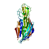
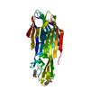
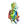
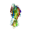
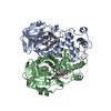
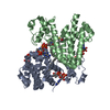
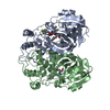
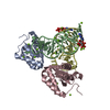
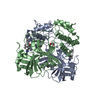
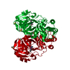
| Entry | Database: PDB / ID: 2qk7 | ||||||
|---|---|---|---|---|---|---|---|
| Title | A covalent S-F heterodimer of staphylococcal gamma-hemolysin | ||||||
 Components Components |
| ||||||
 Keywords Keywords |  TOXIN / pore-forming toxin / beta-barrel / protein-protein interaction / molecular plasticity / covalent complex / Cytolysis / Hemolysis / Secreted TOXIN / pore-forming toxin / beta-barrel / protein-protein interaction / molecular plasticity / covalent complex / Cytolysis / Hemolysis / Secreted | ||||||
| Function / homology |  Function and homology informationcytolysis in another organism / : / toxin activity / extracellular region Function and homology informationcytolysis in another organism / : / toxin activity / extracellular regionSimilarity search - Function | ||||||
| Biological species |   Staphylococcus aureus subsp. aureus (bacteria) Staphylococcus aureus subsp. aureus (bacteria) | ||||||
| Method | X-RAY DIFFRACTION / SYNCHROTRON / MOLECULAR REPLACEMENT / Resolution: 2.4 Å | ||||||
 Authors Authors | Roblin, P. / Guillet, V. / Maveyraud, L. / Mourey, L. | ||||||
 Citation Citation | Journal: Proteins / Year: 2008 Title: A covalent S-F heterodimer of leucotoxin reveals molecular plasticity of beta-barrel pore-forming toxins. Authors: Roblin, P. / Guillet, V. / Joubert, O. / Keller, D. / Erard, M. / Maveyraud, L. / Prevost, G. / Mourey, L. #1: Journal: BIOCHEM.J. / Year: 2006 Title: Engineered covalent leucotoxin heterodimers form functional pores: insights into S-F interactions Authors: Joubert, O. / Viero, G. / Keller, D. / Martinez, E. / Colin, D.A. / Monteil, H. / Mourey, L. / Dalla Serra, M. / Prevost, G. | ||||||
| History |
| ||||||
| Remark 999 | sequence Author stated that these residues are conflicts between different sequence references. |
- Structure visualization
| Structure viewer | Molecule: MolmilJmol/JSmol |
|---|
- Downloads & links
-Download
| PDBx/mmCIF format | 2qk7.cif.gz | 127.1 KB | Display | PDBx/mmCIF format |
|---|---|---|---|---|
| PDB format | pdb2qk7.ent.gz | 96.9 KB | Display | PDB format |
| PDBx/mmJSON format | 2qk7.json.gz | Tree view | PDBx/mmJSON format | |
| Others |  Other downloads Other downloads |
-Validation report
| Arichive directory | https://data.pdbj.org/pub/pdb/validation_reports/qk/2qk7ftp://data.pdbj.org/pub/pdb/validation_reports/qk/2qk7 | HTTPS FTP |
|---|
-Related structure data


-Links
 PDBj
PDBj
- Assembly
| Deposited unit | 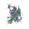
| ||||||||
|---|---|---|---|---|---|---|---|---|---|
| 1 |
| ||||||||
| Unit cell |
| ||||||||
| Details | The disulfide linked gamma-hemolysin heterodimer was prepared from variants HlgA T28C and HlgB N156C as described in Joubert et al., Biochem. J. (2006) 396, 381-389. The asymmetric unit contains the heterodimer. |
-Components
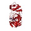
| #1: Protein | Mass: 32725.838 Da / Num. of mol.: 1 / Mutation: T57C Source method: isolated from a genetically manipulated source Source: (gene. exp.) Staphylococcus aureus subsp. aureus (bacteria)Species: Staphylococcus aureus / Strain: MW2 / Gene: hlgA, hlg2 / Plasmid: pGEX-6P-1 / Species (production host): Escherichia coli / Production host: Escherichia coli BL21 (bacteria) / Strain (production host): BL21 / References: UniProt: P0A074 |
|---|---|
| #2: Protein | Mass: 34880.570 Da / Num. of mol.: 1 / Mutation: N182C Source method: isolated from a genetically manipulated source Source: (gene. exp.) Staphylococcus aureus subsp. aureus (bacteria)Species: Staphylococcus aureus / Strain: MW2 / Gene: hlgB / Plasmid: pGEX-6P-1 / Species (production host): Escherichia coli / Production host: Escherichia coli BL21 (bacteria) / Strain (production host): BL21 / References: UniProt: P0A077, UniProt: Q57227*PLUS |
| #3: Water | ChemComp-HOH / Water Mass: 18.015 Da / Num. of mol.: 173 / Source method: isolated from a natural source / Formula: H2O Mass: 18.015 Da / Num. of mol.: 173 / Source method: isolated from a natural source / Formula: H2O |
-Experimental details
-Experiment
| Experiment | Method: X-RAY DIFFRACTION / Number of used crystals: 1 |
|---|
- Sample preparation
| Crystal | Density Matthews: 3.06 Å3/Da / Density % sol: 59.78 % |
|---|---|
| Crystal grow | Temperature: 285 K / Method: vapor diffusion, sitting drop / pH: 5.5 Details: covalent complex concentrated to 9 mg/ml in 50 mM MES-NaOH, 50 mM NaCl, pH 6.5. Needle and rod shape like crystals obtained in drops prepared by mixing two volumes of protein and one volume ...Details: covalent complex concentrated to 9 mg/ml in 50 mM MES-NaOH, 50 mM NaCl, pH 6.5. Needle and rod shape like crystals obtained in drops prepared by mixing two volumes of protein and one volume of reservoir solution containing 10% polyethylene glycol 8000, 0.05 M Na citrate, pH 5.5. Reservoir volumes of 500 mul used, VAPOR DIFFUSION, SITTING DROP, temperature 285K |
-Data collection
| Diffraction | Mean temperature: 100 K |
|---|---|
| Diffraction source | Source: SYNCHROTRON / Site: ESRF  / Beamline: ID14-2 / Wavelength: 0.933 Å / Beamline: ID14-2 / Wavelength: 0.933 Å |
| Detector | Date: Feb 16, 2005 |
| Radiation | Monochromator: Diamond (111), Ge(220) monochromator Sagitally focusing Ge(220) and a multilayer Protocol: SINGLE WAVELENGTH / Monochromatic (M) / Laue (L): M / Scattering type: x-ray |
| Radiation wavelength | Wavelength: 0.933 Å / Relative weight: 1 |
| Reflection | Resolution: 2.4→50.5 Å / Num. all: 32122 / Num. obs: 32122 / % possible obs: 100 % / Redundancy: 7 % / Rsym value: 0.078 / Net I/σ(I): 8 |
| Reflection shell | Resolution: 2.4→2.53 Å / Redundancy: 6.6 % / Mean I/σ(I) obs: 2.3 / Rsym value: 0.318 / % possible all: 100 |
- Processing
| Software |
| ||||||||||||||||||||||||||||||||||||||||||||||||||||||||||||||||||||||||||||||||||||||||||||||||||||||||||||||||||||||||||||||||||||||||||||||||||||||||||||||||||||||||||
|---|---|---|---|---|---|---|---|---|---|---|---|---|---|---|---|---|---|---|---|---|---|---|---|---|---|---|---|---|---|---|---|---|---|---|---|---|---|---|---|---|---|---|---|---|---|---|---|---|---|---|---|---|---|---|---|---|---|---|---|---|---|---|---|---|---|---|---|---|---|---|---|---|---|---|---|---|---|---|---|---|---|---|---|---|---|---|---|---|---|---|---|---|---|---|---|---|---|---|---|---|---|---|---|---|---|---|---|---|---|---|---|---|---|---|---|---|---|---|---|---|---|---|---|---|---|---|---|---|---|---|---|---|---|---|---|---|---|---|---|---|---|---|---|---|---|---|---|---|---|---|---|---|---|---|---|---|---|---|---|---|---|---|---|---|---|---|---|---|---|---|---|
| Refinement | Method to determine structure: MOLECULAR REPLACEMENT Starting model: pdb entries 1LKF, 1T5R Resolution: 2.4→30 Å / Cor.coef. Fo:Fc: 0.946 / Cor.coef. Fo:Fc free: 0.903 / SU B: 6.109 / SU ML: 0.147 / Cross valid method: THROUGHOUT / ESU R: 0.262 / ESU R Free: 0.224 / Stereochemistry target values: MAXIMUM LIKELIHOOD / Details: HYDROGENS HAVE BEEN ADDED IN THE RIDING POSITIONS
| ||||||||||||||||||||||||||||||||||||||||||||||||||||||||||||||||||||||||||||||||||||||||||||||||||||||||||||||||||||||||||||||||||||||||||||||||||||||||||||||||||||||||||
| Solvent computation | Ion probe radii: 0.8 Å / Shrinkage radii: 0.8 Å / VDW probe radii: 1.2 Å / Solvent model: BABINET MODEL WITH MASK | ||||||||||||||||||||||||||||||||||||||||||||||||||||||||||||||||||||||||||||||||||||||||||||||||||||||||||||||||||||||||||||||||||||||||||||||||||||||||||||||||||||||||||
| Displacement parameters | Biso mean: 46.121 Å2
| ||||||||||||||||||||||||||||||||||||||||||||||||||||||||||||||||||||||||||||||||||||||||||||||||||||||||||||||||||||||||||||||||||||||||||||||||||||||||||||||||||||||||||
| Refinement step | Cycle: LAST / Resolution: 2.4→30 Å
| ||||||||||||||||||||||||||||||||||||||||||||||||||||||||||||||||||||||||||||||||||||||||||||||||||||||||||||||||||||||||||||||||||||||||||||||||||||||||||||||||||||||||||
| Refine LS restraints |
| ||||||||||||||||||||||||||||||||||||||||||||||||||||||||||||||||||||||||||||||||||||||||||||||||||||||||||||||||||||||||||||||||||||||||||||||||||||||||||||||||||||||||||
| LS refinement shell | Resolution: 2.4→2.462 Å / Total num. of bins used: 20
|
