 Movie
Movie Controller

[English] 日本語
 Yorodumi
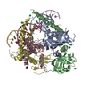
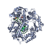
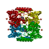
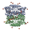
- PDB-2qjs: Stenotrophomonas maltophilia L1 metallo-beta-lactamase Asp-120 As... -
+ Open data

- Basic information
| Entry | Database: PDB / ID: 2qjs | ||||||
|---|---|---|---|---|---|---|---|
| Title | Stenotrophomonas maltophilia L1 metallo-beta-lactamase Asp-120 Asn mutant | ||||||
 Components Components | Metallo-beta-lactamase L1 | ||||||
 Keywords Keywords |  HYDROLASE / metallo-beta-lactamase / dinculear / binculear HYDROLASE / metallo-beta-lactamase / dinculear / binculear | ||||||
| Function / homology |  Function and homology information Function and homology informationbeta-lactam antibiotic catabolic process / antibiotic catabolic process / beta-lactamase activity / beta-lactamase / periplasmic space / response to antibiotic / zinc ion bindingSimilarity search - Function | ||||||
| Biological species |  Stenotrophomonas maltophilia (bacteria) Stenotrophomonas maltophilia (bacteria) | ||||||
| Method | X-RAY DIFFRACTION / SYNCHROTRON / MOLECULAR REPLACEMENT / Resolution: 2.25 Å | ||||||
 Authors Authors | Crisp, J. / Conners, R. / Spencer, J. | ||||||
 Citation Citation | Journal: Biochemistry / Year: 2007 Title: Structural Basis for the Role of Asp-120 in Metallo-beta-lactamases Authors: Crisp, J. / Conners, R. / Garrity, J.D. / Carenbauer, A.L. / Crowder, M.W. / Spencer, J. #1: Journal: J.Biol.Chem. / Year: 2004 Title: Metal Binding Asp-120 in Metallo-beta-lactamase L1 from Stenotrophomonas maltophilia Plays a Crucial Role in Catalysis Authors: Garrity, J.D. / Carenbauer, A.L. / Herron, L.R. / Crowder, M.W. | ||||||
| History |
|
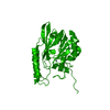
- Structure visualization
| Structure viewer | Molecule: MolmilJmol/JSmol |
|---|
- Downloads & links
-Download
| PDBx/mmCIF format | 2qjs.cif.gz | 205.8 KB | Display | PDBx/mmCIF format |
|---|---|---|---|---|
| PDB format | pdb2qjs.ent.gz | 163.7 KB | Display | PDB format |
| PDBx/mmJSON format | 2qjs.json.gz | Tree view | PDBx/mmJSON format | |
| Others |  Other downloads Other downloads |
-Validation report
| Arichive directory | https://data.pdbj.org/pub/pdb/validation_reports/qj/2qjsftp://data.pdbj.org/pub/pdb/validation_reports/qj/2qjs | HTTPS FTP |
|---|
-Related structure data
| Related structure data |  2qinC  1smlS S: Starting model for refinement C: citing same article ( |
|---|---|
| Similar structure data |


-Links
 PDBj
PDBj

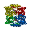
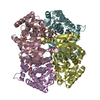
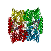
- Assembly
| Deposited unit | 
| ||||||||
|---|---|---|---|---|---|---|---|---|---|
| 1 |
| ||||||||
| 2 | 
| ||||||||
| 3 | 
| ||||||||
| 4 | 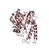
| ||||||||
| 5 | 
| ||||||||
| Unit cell |
|
-Components
| #1: Protein | Mass: 28739.469 Da / Num. of mol.: 4 / Mutation: D120N Source method: isolated from a genetically manipulated source Source: (gene. exp.) Stenotrophomonas maltophilia (bacteria)Strain: IID 1275 / Gene: L1 / Plasmid: pUB5832 / Production host: Escherichia coli (E. coli) / Strain (production host): BL21 (DE3) pLysS / References: UniProt: P52700, beta-lactamase#2: Chemical | ChemComp-ZN /   Mass: 65.409 Da / Num. of mol.: 8 / Source method: obtained synthetically / Formula: Zn Mass: 65.409 Da / Num. of mol.: 8 / Source method: obtained synthetically / Formula: Zn#3: Water | ChemComp-HOH / | Water Mass: 18.015 Da / Num. of mol.: 396 / Source method: isolated from a natural source / Formula: H2O Mass: 18.015 Da / Num. of mol.: 396 / Source method: isolated from a natural source / Formula: H2O |
|---|
-Experimental details
-Experiment
| Experiment | Method: X-RAY DIFFRACTION / Number of used crystals: 1 |
|---|
- Sample preparation
| Crystal | Density Matthews: 2.13 Å3/Da / Density % sol: 42.26 % |
|---|---|
| Crystal grow | Temperature: 293 K / Method: vapor diffusion, hanging drop / pH: 8 Details: 0.1M Tris.Cl pH 8.0, 0.4M MgCl2, 21% PEG 4000, temperature 293K, VAPOR DIFFUSION, HANGING DROP |
-Data collection
| Diffraction | Mean temperature: 100 K |
|---|---|
| Diffraction source | Source: SYNCHROTRON / Site: SRS  / Beamline: PX14.1 / Wavelength: 1.488 Å / Beamline: PX14.1 / Wavelength: 1.488 Å |
| Detector | Type: ADSC QUANTUM 4 / Detector: CCD / Date: Sep 9, 2003 |
| Radiation | Protocol: SINGLE WAVELENGTH / Monochromatic (M) / Laue (L): M / Scattering type: x-ray |
| Radiation wavelength | Wavelength: 1.488 Å / Relative weight: 1 |
| Reflection | Resolution: 2.25→50 Å / Num. all: 47690 / Num. obs: 47129 / % possible obs: 98.8 % / Redundancy: 4.4 % / Biso Wilson estimate: 52.856 Å2 / Rmerge(I) obs: 0.061 / Χ2: 1.026 / Net I/σ(I): 12.3 |
| Reflection shell | Resolution: 2.25→2.33 Å / Redundancy: 4.1 % / Rmerge(I) obs: 0.517 / Mean I/σ(I) obs: 2.3 / Num. unique all: 4559 / Χ2: 1.025 / % possible all: 97.1 |
- Processing
| Software |
| ||||||||||||||||||||||||||||||||||||||||
|---|---|---|---|---|---|---|---|---|---|---|---|---|---|---|---|---|---|---|---|---|---|---|---|---|---|---|---|---|---|---|---|---|---|---|---|---|---|---|---|---|---|
| Refinement | Method to determine structure: MOLECULAR REPLACEMENT Starting model: PDB entry 1SML Resolution: 2.25→10 Å / Num. parameters: 31284 / Num. restraintsaints: 31022 / Cross valid method: FREE R / σ(F): 0 / Stereochemistry target values: ENGH AND HUBER
| ||||||||||||||||||||||||||||||||||||||||
| Refine analyze | Num. disordered residues: 0 / Occupancy sum hydrogen: 0 / Occupancy sum non hydrogen: 7810 | ||||||||||||||||||||||||||||||||||||||||
| Refinement step | Cycle: LAST / Resolution: 2.25→10 Å
| ||||||||||||||||||||||||||||||||||||||||
| Refine LS restraints |
|
