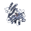
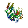
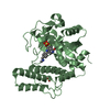
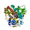
DNA damage-inducible protein DinB / DinB family / dinb family like domain / DinB/YfiT-like putative metalloenzymes / Four Helix Bundle (Hemerythrin (Met), subunit A) / Up-down Bundle / Mainly Alpha Similarity search - Domain/homology
BIOMOLECULE: 1 THIS ENTRY CONTAINS THE CRYSTALLOGRAPHIC ASYMMETRIC UNIT WHICH CONSISTS OF 2 CHAINS. ... BIOMOLECULE: 1 THIS ENTRY CONTAINS THE CRYSTALLOGRAPHIC ASYMMETRIC UNIT WHICH CONSISTS OF 2 CHAINS. SEE REMARK 350 FOR INFORMATION ON GENERATING THE BIOLOGICAL MOLECULE(S). SIZE EXCLUSION CHROMATOGRAPHY SUPPORTS THE ASSIGNMENT OF A DIMER AS THE SIGNIFICANT OLIGOMERIZATION STATE.
Remark 999
SEQUENCE THE CONSTRUCT WAS EXPRESSED WITH A PURIFICATION TAG MGSDKIHHHHHHENLYFQG.
Resolution: 1.9→29.761 Å / Num. obs: 29798 / % possible obs: 100 % / Redundancy: 6.7 % / Rmerge(I) obs: 0.094 / Rsym value: 0.094 / Net I/σ(I): 5.4
Reflection shell
Diffraction-ID: 1
Resolution (Å)
Redundancy (%)
Rmerge(I) obs
Mean I/σ(I) obs
Num. measured all
Num. unique all
Rsym value
% possible all
1.9-1.95
6.9
0.92
0.8
14662
2129
0.92
100
1.95-2
6.8
0.722
1.1
14159
2072
0.722
100
2-2.06
6.8
0.539
1.3
13920
2053
0.539
100
2.06-2.12
6.8
0.41
1.8
13505
1995
0.41
100
2.12-2.19
6.9
0.326
2.3
13080
1905
0.326
100
2.19-2.27
6.8
0.265
2.8
12726
1873
0.265
100
2.27-2.36
6.8
0.231
3.3
12145
1799
0.231
100
2.36-2.45
6.8
0.188
4
11855
1741
0.188
100
2.45-2.56
6.7
0.163
4.6
11368
1688
0.163
100
2.56-2.69
6.7
0.132
5.5
10785
1612
0.132
100
2.69-2.83
6.7
0.117
6.1
10326
1533
0.117
100
2.83-3
6.7
0.096
7.4
9760
1462
0.096
100
3-3.21
6.6
0.088
7.6
9131
1374
0.088
100
3.21-3.47
6.6
0.083
7.7
8518
1298
0.083
100
3.47-3.8
6.4
0.069
8.2
7838
1216
0.069
100
3.8-4.25
6.5
0.058
10.3
7069
1086
0.058
100
4.25-4.91
6.3
0.053
10.8
6268
988
0.053
100
4.91-6.01
6.3
0.05
11.8
5349
855
0.05
100
6.01-8.5
6
0.048
12
4132
693
0.048
100
8.5-29.761
5.1
0.046
12.4
2183
426
0.046
97.6
-
Phasing
Phasing
Method: MAD
-
Processing
Software
Name
Version
Classification
NB
REFMAC
5.2.0019
refinement
PHENIX
refinement
SHELX
phasing
MolProbity
3beta29
modelbuilding
SCALA
datascaling
PDB_EXTRACT
3
dataextraction
MAR345
CCD
datacollection
MOSFLM
datareduction
SHELXD
phasing
SHARP
phasing
Refinement
Method to determine structure: MAD / Resolution: 1.9→29.761 Å / Cor.coef. Fo:Fc: 0.96 / Cor.coef. Fo:Fc free: 0.933 / SU B: 6.23 / SU ML: 0.1 / TLS residual ADP flag: LIKELY RESIDUAL / Cross valid method: THROUGHOUT / σ(F): 0 / ESU R: 0.155 / ESU R Free: 0.143 Stereochemistry target values: MAXIMUM LIKELIHOOD WITH PHASES Details: 1. HYDROGENS HAVE BEEN ADDED IN THE RIDING POSITIONS. 2. A MET-INHIBITION PROTOCOL WAS USED FOR SELENOMETHIONINE INCORPORATION DURING PROTEIN EXPRESSION. THE OCCUPANCY OF THE SE ATOMS IN THE ...Details: 1. HYDROGENS HAVE BEEN ADDED IN THE RIDING POSITIONS. 2. A MET-INHIBITION PROTOCOL WAS USED FOR SELENOMETHIONINE INCORPORATION DURING PROTEIN EXPRESSION. THE OCCUPANCY OF THE SE ATOMS IN THE MSE RESIDUES WAS REDUCED TO 0.75 FOR THE REDUCED SCATTERING POWER DUE TO PARTIAL S-MET INCORPORATION. 3. RESIDUES 94-99 IN CHAIN A AND 152-159 IN CHAIN B ARE DISORDERED AND NOT INCLUDED IN THE MODEL. 4. NI ION IS MODELED BASED ON METAL EXCITATION SCAN. 5. CITRATE ION AND ETHYLENE GLYCOL FROM THE CRYSTALLIZATION/CRYO SOLUTIONS ARE MODELED. 6. ATOM RECORDS CONTAIN RESIDUAL B FACTORS ONLY. 7. RAMACHANDRAN OUTLIER RESIDUE -7 IN CHAIN A LIES IN A DISORDERED REGION. 8. 36 UNUSUALLY STRONG REFLECTIONS WERE OMITTED FROM THE FINAL REFINEMENT.
Rfactor
Num. reflection
% reflection
Selection details
Rfree
0.226
1500
5.1 %
RANDOM
Rwork
0.185
-
-
-
obs
0.187
29655
99.83 %
-
Solvent computation
Ion probe radii: 0.8 Å / Shrinkage radii: 0.8 Å / VDW probe radii: 1.2 Å / Solvent model: BABINET MODEL WITH MASK
Displacement parameters
Biso mean: 27.46 Å2
Baniso -1
Baniso -2
Baniso -3
1-
-1.3 Å2
0 Å2
0 Å2
2-
-
-1.3 Å2
0 Å2
3-
-
-
2.6 Å2
Refinement step
Cycle: LAST / Resolution: 1.9→29.761 Å
Protein
Nucleic acid
Ligand
Solvent
Total
Num. atoms
2716
0
32
235
2983
Refine LS restraints
Refine-ID
Type
Dev ideal
Dev ideal target
Number
X-RAY DIFFRACTION
r_bond_refined_d
0.013
0.021
2853
X-RAY DIFFRACTION
r_bond_other_d
0.004
0.02
1855
X-RAY DIFFRACTION
r_angle_refined_deg
1.429
1.908
3886
X-RAY DIFFRACTION
r_angle_other_deg
1.31
3
4495
X-RAY DIFFRACTION
r_dihedral_angle_1_deg
3.604
5
332
X-RAY DIFFRACTION
r_dihedral_angle_2_deg
32.149
23.514
148
X-RAY DIFFRACTION
r_dihedral_angle_3_deg
10.904
15
465
X-RAY DIFFRACTION
r_dihedral_angle_4_deg
13.224
15
15
X-RAY DIFFRACTION
r_chiral_restr
0.075
0.2
421
X-RAY DIFFRACTION
r_gen_planes_refined
0.006
0.02
3158
X-RAY DIFFRACTION
r_gen_planes_other
0.002
0.02
625
X-RAY DIFFRACTION
r_nbd_refined
0.188
0.2
630
X-RAY DIFFRACTION
r_nbd_other
0.139
0.2
1860
X-RAY DIFFRACTION
r_nbtor_refined
0.167
0.2
1377
X-RAY DIFFRACTION
r_nbtor_other
0.072
0.2
1322
X-RAY DIFFRACTION
r_xyhbond_nbd_refined
0.099
0.2
196
X-RAY DIFFRACTION
r_symmetry_vdw_refined
0.179
0.2
10
X-RAY DIFFRACTION
r_symmetry_vdw_other
0.118
0.2
35
X-RAY DIFFRACTION
r_symmetry_hbond_refined
0.085
0.2
13
X-RAY DIFFRACTION
r_mcbond_it
1.733
3
1724
X-RAY DIFFRACTION
r_mcbond_other
0.424
3
665
X-RAY DIFFRACTION
r_mcangle_it
2.608
5
2658
X-RAY DIFFRACTION
r_scbond_it
4.402
8
1361
X-RAY DIFFRACTION
r_scangle_it
5.67
11
1224
LS refinement shell
Resolution: 1.9→1.949 Å / Total num. of bins used: 20
Rfactor
Num. reflection
% reflection
Rfree
0.296
103
-
Rwork
0.232
2019
-
obs
-
2122
99.86 %
Refinement TLS params.
Method: refined / Refine-ID: X-RAY DIFFRACTION
ID
L11 (°2)
L12 (°2)
L13 (°2)
L22 (°2)
L23 (°2)
L33 (°2)
S11 (Å °)
S12 (Å °)
S13 (Å °)
S21 (Å °)
S22 (Å °)
S23 (Å °)
S31 (Å °)
S32 (Å °)
S33 (Å °)
T11 (Å2)
T12 (Å2)
T13 (Å2)
T22 (Å2)
T23 (Å2)
T33 (Å2)
Origin x (Å)
Origin y (Å)
Origin z (Å)
1
1.9003
-0.874
0.0972
2.6665
-0.399
1.8746
0.1346
0.3449
0.016
-0.2775
-0.2076
-0.1769
0.0833
0.1811
0.073
-0.1418
0.0611
0.0159
-0.0883
-0.0025
-0.276
-10.638
17.651
11.831
2
2.0169
0.5682
0.7184
2.1154
0.4827
1.7709
0.0934
0.0234
0.0525
0.0816
-0.0829
0.3351
-0.0101
-0.2533
-0.0105
-0.1604
0.0216
0.0228
-0.141
-0.025
-0.163
-29.089
24.314
22.265
Refinement TLS group
ID
Refine-ID
Refine TLS-ID
Auth asym-ID
Label asym-ID
Auth seq-ID
Label seq-ID
1
X-RAY DIFFRACTION
1
A
A
-11 - 93
8 - 112
2
X-RAY DIFFRACTION
1
A
A
100 - 159
119 - 178
3
X-RAY DIFFRACTION
2
B
B
-10 - 151
9 - 170
+
About Yorodumi
-
News
-
Feb 9, 2022. New format data for meta-information of EMDB entries
New format data for meta-information of EMDB entries
Version 3 of the EMDB header file is now the official format.
The previous official version 1.9 will be removed from the archive.
In the structure databanks used in Yorodumi, some data are registered as the other names, "COVID-19 virus" and "2019-nCoV". Here are the details of the virus and the list of structure data.
Jan 31, 2019. EMDB accession codes are about to change! (news from PDBe EMDB page)
EMDB accession codes are about to change! (news from PDBe EMDB page)
The allocation of 4 digits for EMDB accession codes will soon come to an end. Whilst these codes will remain in use, new EMDB accession codes will include an additional digit and will expand incrementally as the available range of codes is exhausted. The current 4-digit format prefixed with “EMD-” (i.e. EMD-XXXX) will advance to a 5-digit format (i.e. EMD-XXXXX), and so on. It is currently estimated that the 4-digit codes will be depleted around Spring 2019, at which point the 5-digit format will come into force.
The EM Navigator/Yorodumi systems omit the EMD- prefix.
Related info.:Q: What is EMD? / ID/Accession-code notation in Yorodumi/EM Navigator
Yorodumi is a browser for structure data from EMDB, PDB, SASBDB, etc.
This page is also the successor to EM Navigator detail page, and also detail information page/front-end page for Omokage search.
The word "yorodu" (or yorozu) is an old Japanese word meaning "ten thousand". "mi" (miru) is to see.
Related info.:EMDB / PDB / SASBDB / Comparison of 3 databanks / Yorodumi Search / Aug 31, 2016. New EM Navigator & Yorodumi / Yorodumi Papers / Jmol/JSmol / Function and homology information / Changes in new EM Navigator and Yorodumi
 Movie
Movie Controller

 Yorodumi
 Open data
 Basic information
 Components
 Keywords
 HYDROLASE / Dinb/yfit-like putative metalloenzymes fold /
  Function and homology information
 Authors
 Citation
 Structure visualization
 Downloads & links
 Other downloads


 PDBj
PDBj Assembly

 Mass: 58.693 Da / Num. of mol.: 2 / Source method: obtained synthetically / Formula: Ni
 Mass: 192.124 Da / Num. of mol.: 2 / Source method: obtained synthetically / Formula: C6H8O7
 Mass: 62.068 Da / Num. of mol.: 1 / Source method: obtained synthetically / Formula: C2H6O2
 Mass: 18.015 Da / Num. of mol.: 235 / Source method: isolated from a natural source / Formula: H2O
 Sample preparation
 / Beamline: BL9-2 / Wavelength: 0.91162, 0.97925
 Processing