 Movie
Movie Controller

[English] 日本語
 Yorodumi
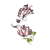
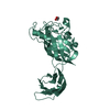
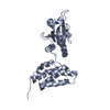
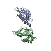
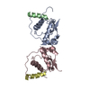
- PDB-2q82: Crystal structure of core protein P7 from Pseudomonas phage phi12... -
+ Open data

- Basic information
| Entry | Database: PDB / ID: 2q82 | ||||||
|---|---|---|---|---|---|---|---|
| Title | Crystal structure of core protein P7 from Pseudomonas phage phi12. Northeast Structural Genomics Target OC1 | ||||||
 Components Components | Core protein P7 | ||||||
 Keywords Keywords |  STRUCTURAL PROTEIN / P7 / core protein / NESG / OC1 / structural genomics / PSI-2 / Protein Structure Initiative / Northeast Structural Genomics Consortium STRUCTURAL PROTEIN / P7 / core protein / NESG / OC1 / structural genomics / PSI-2 / Protein Structure Initiative / Northeast Structural Genomics Consortium | ||||||
| Function / homology | Oxidized Rhodanese; domain 1 - #20 / Oxidized Rhodanese; domain 1 / 3-Layer(aba) Sandwich / Alpha Beta / Core protein P7 Function and homology information Function and homology information | ||||||
| Biological species |  Pseudomonas phage phi12 (bacteriophage) Pseudomonas phage phi12 (bacteriophage) | ||||||
| Method | X-RAY DIFFRACTION / SYNCHROTRON / SAD / Resolution: 1.83 Å | ||||||
 Authors Authors | Benach, J. / Eryilmaz, E. / Su, M. / Seetharaman, J. / Wei, H. / Gottlieb, P. / Hunt, J.F. / Ghose, R. / Northeast Structural Genomics Consortium (NESG) | ||||||
 Citation Citation | Journal: J.Mol.Biol. / Year: 2008 Title: Structure and dynamics of the P7 protein from the bacteriophage phi 12. Authors: Eryilmaz, E. / Benach, J. / Su, M. / Seetharaman, J. / Dutta, K. / Wei, H. / Gottlieb, P. / Hunt, J.F. / Ghose, R. | ||||||
| History |
|
- Structure visualization
| Structure viewer | Molecule: MolmilJmol/JSmol |
|---|
- Downloads & links
-Download
| PDBx/mmCIF format | 2q82.cif.gz | 37.3 KB | Display | PDBx/mmCIF format |
|---|---|---|---|---|
| PDB format | pdb2q82.ent.gz | 26.8 KB | Display | PDB format |
| PDBx/mmJSON format | 2q82.json.gz | Tree view | PDBx/mmJSON format | |
| Others |  Other downloads Other downloads |
-Validation report
| Arichive directory | https://data.pdbj.org/pub/pdb/validation_reports/q8/2q82ftp://data.pdbj.org/pub/pdb/validation_reports/q8/2q82 | HTTPS FTP |
|---|
-Related structure data
| Similar structure data | |
|---|---|
| Other databases |


-Links
 PDBj
PDBj- Assembly
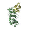
| Deposited unit | 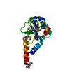
| ||||||||
|---|---|---|---|---|---|---|---|---|---|
| 1 | 
| ||||||||
| Unit cell |
| ||||||||
| Details | The putative biological unit is a dimer, created by -x, -x+y, -z+2/3 + (1 1 0) symmetry operation |
-Components
| #1: Protein | Mass: 14450.371 Da / Num. of mol.: 1 / Fragment: Residues 1-129 Source method: isolated from a genetically manipulated source Source: (gene. exp.) Pseudomonas phage phi12 (bacteriophage)Genus: Cystovirus / Plasmid: pET21D / Production host:  Escherichia coli (E. coli) / Strain (production host): B834(DE3) / References: UniProt: Q94M07 Escherichia coli (E. coli) / Strain (production host): B834(DE3) / References: UniProt: Q94M07 |
|---|---|
| #2: Water | ChemComp-HOH / Water Mass: 18.015 Da / Num. of mol.: 188 / Source method: isolated from a natural source / Formula: H2O Mass: 18.015 Da / Num. of mol.: 188 / Source method: isolated from a natural source / Formula: H2O |
-Experimental details
-Experiment
| Experiment | Method: X-RAY DIFFRACTION / Number of used crystals: 1 |
|---|
- Sample preparation
| Crystal | Density Matthews: 2.65 Å3/Da / Density % sol: 53.56 % Description: THE STRUCTURE FACTOR FILE CONTAINS FRIEDEL PAIRS |
|---|---|
| Crystal grow | Temperature: 293 K / Method: vapor diffusion, hanging drop Details: 23% PEG10000, 100mM Sodium acetate, 10% n-Hexyl-D-glucoside, VAPOR DIFFUSION, HANGING DROP, temperature 293K |
-Data collection
| Diffraction | Mean temperature: 100 K | |||||||||||||||||||||||||||||||||||||||||||||||||||||||||||||||||||||||||||||
|---|---|---|---|---|---|---|---|---|---|---|---|---|---|---|---|---|---|---|---|---|---|---|---|---|---|---|---|---|---|---|---|---|---|---|---|---|---|---|---|---|---|---|---|---|---|---|---|---|---|---|---|---|---|---|---|---|---|---|---|---|---|---|---|---|---|---|---|---|---|---|---|---|---|---|---|---|---|---|
| Diffraction source | Source: SYNCHROTRON / Site: NSLS  / Beamline: X12B / Wavelength: 0.97947 Å / Beamline: X12B / Wavelength: 0.97947 Å | |||||||||||||||||||||||||||||||||||||||||||||||||||||||||||||||||||||||||||||
| Detector | Type: MAR CCD 165 mm / Detector: CCD / Date: May 8, 2007 / Details: mirrors | |||||||||||||||||||||||||||||||||||||||||||||||||||||||||||||||||||||||||||||
| Radiation | Monochromator: SI(111) / Protocol: SINGLE WAVELENGTH / Monochromatic (M) / Laue (L): M / Scattering type: x-ray | |||||||||||||||||||||||||||||||||||||||||||||||||||||||||||||||||||||||||||||
| Radiation wavelength | Wavelength: 0.97947 Å / Relative weight: 1 | |||||||||||||||||||||||||||||||||||||||||||||||||||||||||||||||||||||||||||||
| Reflection | Redundancy: 10.5 % / Av σ(I) over netI: 12 / Number: 305699 / Rmerge(I) obs: 0.05 / Χ2: 0.78 / D res high: 1.7 Å / D res low: 50 Å / Num. obs: 29155 / % possible obs: 89.4 | |||||||||||||||||||||||||||||||||||||||||||||||||||||||||||||||||||||||||||||
| Diffraction reflection shell |
| |||||||||||||||||||||||||||||||||||||||||||||||||||||||||||||||||||||||||||||
| Reflection | Resolution: 1.7→50 Å / Num. obs: 29155 / % possible obs: 89.4 % / Observed criterion σ(I): -3 / Redundancy: 10.5 % / Rmerge(I) obs: 0.05 / Net I/σ(I): 12 | |||||||||||||||||||||||||||||||||||||||||||||||||||||||||||||||||||||||||||||
| Reflection shell |
|
-Phasing
| Phasing | Method: SAD | |||||||||||||||||||||||||||||||||||||||||||||||||
|---|---|---|---|---|---|---|---|---|---|---|---|---|---|---|---|---|---|---|---|---|---|---|---|---|---|---|---|---|---|---|---|---|---|---|---|---|---|---|---|---|---|---|---|---|---|---|---|---|---|---|
| Phasing MAD | D res high: 1.88 Å / D res low: 5.83 Å / FOM : 0.295 / Reflection: 9638 | |||||||||||||||||||||||||||||||||||||||||||||||||
| Phasing MAD set | R kraut acentric: 0.037 / Highest resolution: 5.83 Å / Lowest resolution: 1.88 Å / FOM : 0.297 / Power: 0.71 kW | |||||||||||||||||||||||||||||||||||||||||||||||||
| Phasing dm | FOM : 0.52 / FOM acentric: 0.52 / FOM centric: 0.5 / Reflection: 12613 / Reflection acentric: 11383 / Reflection centric: 1230 | |||||||||||||||||||||||||||||||||||||||||||||||||
| Phasing dm shell |
|
- Processing
| Software |
| ||||||||||||||||||||||||||||||||||||||||||||||||||||||||||||||||||||||||||||||||||||||||||||||||||||||||||||||||||||||||||||||||||||||||||||||||||||||||||||||||||||||||||||||||||||||||||||||||||||||||||||||||||||||||||||||||||||||||||||||||||||||||||||||||||||||||||||||||||||||||||||||||||||||
|---|---|---|---|---|---|---|---|---|---|---|---|---|---|---|---|---|---|---|---|---|---|---|---|---|---|---|---|---|---|---|---|---|---|---|---|---|---|---|---|---|---|---|---|---|---|---|---|---|---|---|---|---|---|---|---|---|---|---|---|---|---|---|---|---|---|---|---|---|---|---|---|---|---|---|---|---|---|---|---|---|---|---|---|---|---|---|---|---|---|---|---|---|---|---|---|---|---|---|---|---|---|---|---|---|---|---|---|---|---|---|---|---|---|---|---|---|---|---|---|---|---|---|---|---|---|---|---|---|---|---|---|---|---|---|---|---|---|---|---|---|---|---|---|---|---|---|---|---|---|---|---|---|---|---|---|---|---|---|---|---|---|---|---|---|---|---|---|---|---|---|---|---|---|---|---|---|---|---|---|---|---|---|---|---|---|---|---|---|---|---|---|---|---|---|---|---|---|---|---|---|---|---|---|---|---|---|---|---|---|---|---|---|---|---|---|---|---|---|---|---|---|---|---|---|---|---|---|---|---|---|---|---|---|---|---|---|---|---|---|---|---|---|---|---|---|---|---|---|---|---|---|---|---|---|---|---|---|---|---|---|---|---|---|---|---|---|---|---|---|---|---|---|---|---|---|---|---|---|---|---|---|---|---|---|---|---|---|---|---|---|---|---|---|---|---|
| Refinement | Method to determine structure: SAD / Resolution: 1.83→20 Å / σ(F): 2 / Stereochemistry target values: Engh & Huber / Details: THE FRIEDEL PAIRS WERE USED FOR PHASING
| ||||||||||||||||||||||||||||||||||||||||||||||||||||||||||||||||||||||||||||||||||||||||||||||||||||||||||||||||||||||||||||||||||||||||||||||||||||||||||||||||||||||||||||||||||||||||||||||||||||||||||||||||||||||||||||||||||||||||||||||||||||||||||||||||||||||||||||||||||||||||||||||||||||||
| Solvent computation | Bsol: 52.745 Å2 | ||||||||||||||||||||||||||||||||||||||||||||||||||||||||||||||||||||||||||||||||||||||||||||||||||||||||||||||||||||||||||||||||||||||||||||||||||||||||||||||||||||||||||||||||||||||||||||||||||||||||||||||||||||||||||||||||||||||||||||||||||||||||||||||||||||||||||||||||||||||||||||||||||||||
| Displacement parameters | Biso mean: 39.389 Å2
| ||||||||||||||||||||||||||||||||||||||||||||||||||||||||||||||||||||||||||||||||||||||||||||||||||||||||||||||||||||||||||||||||||||||||||||||||||||||||||||||||||||||||||||||||||||||||||||||||||||||||||||||||||||||||||||||||||||||||||||||||||||||||||||||||||||||||||||||||||||||||||||||||||||||
| Refinement step | Cycle: LAST / Resolution: 1.83→20 Å
| ||||||||||||||||||||||||||||||||||||||||||||||||||||||||||||||||||||||||||||||||||||||||||||||||||||||||||||||||||||||||||||||||||||||||||||||||||||||||||||||||||||||||||||||||||||||||||||||||||||||||||||||||||||||||||||||||||||||||||||||||||||||||||||||||||||||||||||||||||||||||||||||||||||||
| LS refinement shell |
| ||||||||||||||||||||||||||||||||||||||||||||||||||||||||||||||||||||||||||||||||||||||||||||||||||||||||||||||||||||||||||||||||||||||||||||||||||||||||||||||||||||||||||||||||||||||||||||||||||||||||||||||||||||||||||||||||||||||||||||||||||||||||||||||||||||||||||||||||||||||||||||||||||||||
| Xplor file |
|
