 Movie
Movie Controller


+ Open data

- Basic information
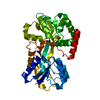
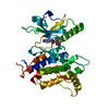
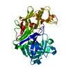
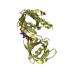
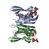
| Entry | Database: PDB / ID: 2q3x | ||||||
|---|---|---|---|---|---|---|---|
| Title | The RIM1alpha C2B domain | ||||||
 Components Components | Regulating synaptic membrane exocytosis protein 1 | ||||||
 Keywords Keywords |  TRANSPORT PROTEIN / C2 DOMAIN DIMER / NEUROTRANSMITTER RELEASE TRANSPORT PROTEIN / C2 DOMAIN DIMER / NEUROTRANSMITTER RELEASE | ||||||
| Function / homology |  Function and homology information Function and homology informationpositive regulation of synaptic vesicle priming / positive regulation of synaptic vesicle fusion to presynaptic active zone membrane / extrinsic component of presynaptic active zone membrane / acrosomal vesicle exocytosis / regulation of calcium-dependent activation of synaptic vesicle fusion / structural constituent of presynaptic active zone / cytoskeleton of presynaptic active zone / spontaneous neurotransmitter secretion / Glutamate Neurotransmitter Release Cycle / Norepinephrine Neurotransmitter Release Cycle ...positive regulation of synaptic vesicle priming / positive regulation of synaptic vesicle fusion to presynaptic active zone membrane / extrinsic component of presynaptic active zone membrane / acrosomal vesicle exocytosis / regulation of calcium-dependent activation of synaptic vesicle fusion / structural constituent of presynaptic active zone / cytoskeleton of presynaptic active zone / spontaneous neurotransmitter secretion / Glutamate Neurotransmitter Release Cycle / Norepinephrine Neurotransmitter Release Cycle / Acetylcholine Neurotransmitter Release Cycle / Serotonin Neurotransmitter Release Cycle / GABA synthesis, release, reuptake and degradation / Dopamine Neurotransmitter Release Cycle / presynaptic dense core vesicle exocytosis / synaptic vesicle docking / inhibitory synapse / calcium ion-regulated exocytosis of neurotransmitter / presynaptic active zone cytoplasmic component / positive regulation of dendrite extension / positive regulation of inhibitory postsynaptic potential / neurotransmitter secretion / synaptic vesicle priming / regulation of synaptic vesicle exocytosis / positive regulation of excitatory postsynaptic potential / GABA-ergic synapse / positive regulation of synaptic transmission / regulation of membrane potential / cell projection / long-term synaptic potentiation / regulation of long-term neuronal synaptic plasticity / intracellular protein transport / regulation of synaptic plasticity / small GTPase binding / SH3 domain binding / presynaptic membrane / protein-containing complex assembly / vesicle / transmembrane transporter binding / postsynaptic density / cell differentiation / glutamatergic synapse / synapse / positive regulation of gene expression / protein kinase binding / metal ion binding / plasma membraneSimilarity search - Function | ||||||
| Biological species |  Rattus norvegicus (Norway rat) Rattus norvegicus (Norway rat) | ||||||
| Method | X-RAY DIFFRACTION / SYNCHROTRON / SAD / Resolution: 1.73 Å | ||||||
 Authors Authors | Guan, R. / Dai, H. / Tomchick, D.R. / Machius, M. / Sudhof, T.C. / Rizo, J. | ||||||
 Citation Citation | Journal: Biochemistry / Year: 2007 Title: Crystal Structure of the RIM1alpha C(2)B Domain at 1.7 A Resolution. Authors: Guan, R. / Dai, H. / Tomchick, D.R. / Dulubova, I. / Machius, M. / Sudhof, T.C. / Rizo, J. #1: Journal: Nature / Year: 2002Title: RIM1alpha forms a protein scaffold for regulating neurotransmitter release at the active zone. Authors: Schoch, S. / Castillo, P.E. / Jo, T. / Mukherjee, K. / Geppert, M. / Wang, Y. / Schmitz, F. / Malenka, R.C. / Sudhof, T.C. #2: Journal: Nature / Year: 2002Title: RIM1alpha is required for presynaptic long-term potentiation. Authors: Castillo, P.E. / Schoch, S. / Schmitz, F. / Sudhof, T.C. / Malenka, R.C. #3: Journal: Neuron / Year: 2004Title: The presynaptic active zone protein RIM1alpha is critical for normal learning and memory. Authors: Powell, C.M. / Schoch, S. / Monteggia, L. / Barrot, M. / Matos, M.F. / Feldmann, N. / Sudhof, T.C. / Nestler, E.J. #4: Journal: Biochem.Soc.Trans. / Year: 2005Title: RIM function in short- and long-term synaptic plasticity. Authors: Kaeser, P.S. / Sudhof, T.C. #5: Journal: J.Biol.Chem. / Year: 1998Title: C2-domains, structure and function of a universal Ca2+-binding domain. Authors: Rizo, J. / Sudhof, T.C. | ||||||
| History |
|
- Structure visualization
| Structure viewer | Molecule: MolmilJmol/JSmol |
|---|
- Downloads & links
-Download
| PDBx/mmCIF format | 2q3x.cif.gz | 71.9 KB | Display | PDBx/mmCIF format |
|---|---|---|---|---|
| PDB format | pdb2q3x.ent.gz | 58.7 KB | Display | PDB format |
| PDBx/mmJSON format | 2q3x.json.gz | Tree view | PDBx/mmJSON format | |
| Others |  Other downloads Other downloads |
-Validation report
| Arichive directory | https://data.pdbj.org/pub/pdb/validation_reports/q3/2q3xftp://data.pdbj.org/pub/pdb/validation_reports/q3/2q3x | HTTPS FTP |
|---|
-Related structure data
| Similar structure data |
|---|


-Links
 PDBj
PDBj


- Assembly
| Deposited unit | 
| ||||||||
|---|---|---|---|---|---|---|---|---|---|
| 1 |
| ||||||||
| Unit cell |
| ||||||||
| Components on special symmetry positions |
| ||||||||
| Details | The contents of the asymmetric unit is a homodimer. |
-Components
| #1: Protein | Mass: 18958.188 Da / Num. of mol.: 2 / Fragment: C2B domain Source method: isolated from a genetically manipulated source Source: (gene. exp.) Rattus norvegicus (Norway rat) / Gene: Rims1, Rim1 / Plasmid: pGEX-KT / Species (production host): Escherichia coli / Production host:  Escherichia coli BL21 (bacteria) / Strain (production host): BL21 / References: UniProt: Q9JIR4 Escherichia coli BL21 (bacteria) / Strain (production host): BL21 / References: UniProt: Q9JIR4#2: Chemical | ChemComp-SO4 / Sulfate  Mass: 96.063 Da / Num. of mol.: 7 / Source method: obtained synthetically / Formula: SO4 Mass: 96.063 Da / Num. of mol.: 7 / Source method: obtained synthetically / Formula: SO4#3: Chemical | Chloride  Mass: 35.453 Da / Num. of mol.: 3 / Source method: obtained synthetically / Formula: Cl Mass: 35.453 Da / Num. of mol.: 3 / Source method: obtained synthetically / Formula: Cl#4: Chemical | ChemComp-NA / |   Mass: 22.990 Da / Num. of mol.: 1 / Source method: obtained synthetically / Formula: Na Mass: 22.990 Da / Num. of mol.: 1 / Source method: obtained synthetically / Formula: Na#5: Water | ChemComp-HOH / | Water Mass: 18.015 Da / Num. of mol.: 189 / Source method: isolated from a natural source / Formula: H2O Mass: 18.015 Da / Num. of mol.: 189 / Source method: isolated from a natural source / Formula: H2O |
|---|
-Experimental details
-Experiment
| Experiment | Method: X-RAY DIFFRACTION / Number of used crystals: 1 |
|---|
- Sample preparation
| Crystal | Density Matthews: 2.13 Å3/Da / Density % sol: 42.13 % |
|---|---|
| Crystal grow | Temperature: 293 K / Method: vapor diffusion, hanging drop / pH: 3.5 Details: 1.9 M ammonium sulfate, 0.1 M sodium citrate, 0.15 M NaCl, 20% ethylene glycol, pH 3.5, VAPOR DIFFUSION, HANGING DROP, temperature 293K |
-Data collection
| Diffraction | Mean temperature: 100 K |
|---|---|
| Diffraction source | Source: SYNCHROTRON / Site: APS  / Beamline: 19-BM / Wavelength: 0.97918 Å / Beamline: 19-BM / Wavelength: 0.97918 Å |
| Detector | Type: ADSC QUANTUM 315 / Detector: CCD / Date: Feb 17, 2006 |
| Radiation | Monochromator: Si 111 / Protocol: SINGLE WAVELENGTH / Monochromatic (M) / Laue (L): M / Scattering type: x-ray |
| Radiation wavelength | Wavelength: 0.97918 Å / Relative weight: 1 |
| Reflection | Resolution: 1.73→26.4 Å / Num. all: 34669 / Num. obs: 34669 / % possible obs: 95.3 % / Observed criterion σ(F): 1 / Observed criterion σ(I): 1 |
| Reflection shell | Resolution: 1.73→1.775 Å / Redundancy: 7.1 % / Rmerge(I) obs: 0.747 / Mean I/σ(I) obs: 2.1 / % possible all: 100 |
- Processing
| Software |
| ||||||||||||||||||||||||||||||||||||||||||||||||||||||||||||||||||||||||||||||||||||||||||
|---|---|---|---|---|---|---|---|---|---|---|---|---|---|---|---|---|---|---|---|---|---|---|---|---|---|---|---|---|---|---|---|---|---|---|---|---|---|---|---|---|---|---|---|---|---|---|---|---|---|---|---|---|---|---|---|---|---|---|---|---|---|---|---|---|---|---|---|---|---|---|---|---|---|---|---|---|---|---|---|---|---|---|---|---|---|---|---|---|---|---|---|
| Refinement | Method to determine structure: SAD / Resolution: 1.73→26.4 Å / Cor.coef. Fo:Fc: 0.963 / Cor.coef. Fo:Fc free: 0.941 / SU B: 3.82 / SU ML: 0.063 / TLS residual ADP flag: LIKELY RESIDUAL / Cross valid method: THROUGHOUT / σ(F): 0 / σ(I): 0 / ESU R: 0.102 / ESU R Free: 0.103 / Stereochemistry target values: MAXIMUM LIKELIHOOD / Details: HYDROGENS HAVE BEEN ADDED IN THE RIDING POSITIONS
| ||||||||||||||||||||||||||||||||||||||||||||||||||||||||||||||||||||||||||||||||||||||||||
| Solvent computation | Ion probe radii: 0.8 Å / Shrinkage radii: 0.8 Å / VDW probe radii: 1.4 Å / Solvent model: MASK | ||||||||||||||||||||||||||||||||||||||||||||||||||||||||||||||||||||||||||||||||||||||||||
| Displacement parameters | Biso mean: 25.991 Å2
| ||||||||||||||||||||||||||||||||||||||||||||||||||||||||||||||||||||||||||||||||||||||||||
| Refine analyze | Luzzati coordinate error obs: 0.21 Å / Luzzati d res low obs: 6 Å | ||||||||||||||||||||||||||||||||||||||||||||||||||||||||||||||||||||||||||||||||||||||||||
| Refinement step | Cycle: LAST / Resolution: 1.73→26.4 Å
| ||||||||||||||||||||||||||||||||||||||||||||||||||||||||||||||||||||||||||||||||||||||||||
| Refine LS restraints |
| ||||||||||||||||||||||||||||||||||||||||||||||||||||||||||||||||||||||||||||||||||||||||||
| LS refinement shell | Resolution: 1.73→1.775 Å / Total num. of bins used: 20
| ||||||||||||||||||||||||||||||||||||||||||||||||||||||||||||||||||||||||||||||||||||||||||
| Refinement TLS params. | Method: refined / Refine-ID: X-RAY DIFFRACTION
| ||||||||||||||||||||||||||||||||||||||||||||||||||||||||||||||||||||||||||||||||||||||||||
| Refinement TLS group |
|
