Group: Derived calculations / Version format compliance
Remark 300
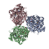
BIOMOLECULE: 1 THIS ENTRY CONTAINS THE CRYSTALLOGRAPHIC ASYMMETRIC UNIT WHICH CONSISTS OF 4 ... BIOMOLECULE: 1 THIS ENTRY CONTAINS THE CRYSTALLOGRAPHIC ASYMMETRIC UNIT WHICH CONSISTS OF 4 CHAIN(S). AUTHORS STATE THAT THE BIOLOGICAL UNIT OF THIS PROTEIN IS EXPERIMENTALLY UNKNOWN. THE DODECAMERIC ASSEMBLY OF THE BIOLOGICAL UNIT, SHOWN IN REMARK 350, IS PREDICTED BY THE ANALYSIS OF PROTEIN INTERFACES (PISA) BASED ON THIS CRYSTAL STRUCTURE.
Remark 999
SEQUENCE AUTHORS STATE THAT THE ELECTRON DENSITY CLEARLY SHOWS THE PRESENCE OF ARGININE AT ... SEQUENCE AUTHORS STATE THAT THE ELECTRON DENSITY CLEARLY SHOWS THE PRESENCE OF ARGININE AT SEQUENCE POSITION 20 IN ALL FOUR CHAINS A-D IN THE ASYMMETRIC UNIT. THE PRESENCE OF ARGININES IS SUPPORTED BY OMIT MAPS AND BY THE R-FACTOR INCREASE WHEN LYSINES ARE BEING REFINED AT THAT POSITION.
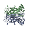
A: Dual specificity protein phosphatase B: Dual specificity protein phosphatase C: Dual specificity protein phosphatase D: Dual specificity protein phosphatase
A: Dual specificity protein phosphatase B: Dual specificity protein phosphatase C: Dual specificity protein phosphatase D: Dual specificity protein phosphatase
A: Dual specificity protein phosphatase B: Dual specificity protein phosphatase C: Dual specificity protein phosphatase D: Dual specificity protein phosphatase
A: Dual specificity protein phosphatase B: Dual specificity protein phosphatase C: Dual specificity protein phosphatase D: Dual specificity protein phosphatase
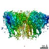
Resolution: 2.57→2.66 Å / Redundancy: 8.8 % / Rmerge(I) obs: 0.899 / Mean I/σ(I) obs: 2.21 / Num. unique all: 4979 / % possible all: 97.7
-
Processing
Software
Name
Version
Classification
REFMAC
5.2.0019
refinement
SBC-Collect
datacollection
HKL-3000
datareduction
HKL-3000
datascaling
SHELXD
phasing
MLPHARE
phasing
DM
phasing
SOLVE
phasing
RESOLVE
phasing
ARP/wARP
modelbuilding
HKL-3000
phasing
Refinement
Method to determine structure: SAD / Resolution: 2.57→40 Å / Cor.coef. Fo:Fc: 0.949 / Cor.coef. Fo:Fc free: 0.93 / SU B: 13.217 / SU ML: 0.137 / Cross valid method: THROUGHOUT / σ(F): 0 / σ(I): 0 / ESU R: 0.24 / ESU R Free: 0.198 / Stereochemistry target values: MAXIMUM LIKELIHOOD Details: HYDROGENS HAVE BEEN ADDED IN THE RIDING POSITIONS. Only fragments of N-terminal parts (His-tags) of chains A and D were identified in the electron density and modeled with the side chain atoms missing.
Rfactor
Num. reflection
% reflection
Selection details
Rfree
0.2183
2610
5.1 %
RANDOM
Rwork
0.1913
-
-
-
all
0.1927
48741
-
-
obs
0.1927
48741
99.67 %
-
Solvent computation
Ion probe radii: 0.8 Å / Shrinkage radii: 0.8 Å / VDW probe radii: 1.2 Å / Solvent model: MASK
Displacement parameters
Biso mean: 35.961 Å2
Refinement step
Cycle: LAST / Resolution: 2.57→40 Å
Protein
Nucleic acid
Ligand
Solvent
Total
Num. atoms
5738
0
0
63
5801
Refine LS restraints
Refine-ID
Type
Dev ideal
Dev ideal target
Number
X-RAY DIFFRACTION
r_bond_refined_d
0.016
0.022
5887
X-RAY DIFFRACTION
r_bond_other_d
X-RAY DIFFRACTION
r_angle_refined_deg
1.523
1.966
7951
X-RAY DIFFRACTION
r_angle_other_deg
X-RAY DIFFRACTION
r_dihedral_angle_1_deg
5.811
5
715
X-RAY DIFFRACTION
r_dihedral_angle_2_deg
38.168
23.779
262
X-RAY DIFFRACTION
r_dihedral_angle_3_deg
19.445
15
1081
X-RAY DIFFRACTION
r_dihedral_angle_4_deg
19.885
15
32
X-RAY DIFFRACTION
r_chiral_restr
0.11
0.2
888
X-RAY DIFFRACTION
r_gen_planes_refined
0.005
0.02
4361
X-RAY DIFFRACTION
r_gen_planes_other
X-RAY DIFFRACTION
r_nbd_refined
0.226
0.2
2561
X-RAY DIFFRACTION
r_nbd_other
X-RAY DIFFRACTION
r_nbtor_refined
0.321
0.2
4003
X-RAY DIFFRACTION
r_nbtor_other
X-RAY DIFFRACTION
r_xyhbond_nbd_refined
0.129
0.2
216
X-RAY DIFFRACTION
r_xyhbond_nbd_other
X-RAY DIFFRACTION
r_metal_ion_refined
X-RAY DIFFRACTION
r_metal_ion_other
X-RAY DIFFRACTION
r_symmetry_vdw_refined
0.263
0.2
74
X-RAY DIFFRACTION
r_symmetry_vdw_other
X-RAY DIFFRACTION
r_symmetry_hbond_refined
0.091
0.2
8
X-RAY DIFFRACTION
r_symmetry_hbond_other
X-RAY DIFFRACTION
r_symmetry_metal_ion_refined
X-RAY DIFFRACTION
r_symmetry_metal_ion_other
X-RAY DIFFRACTION
r_mcbond_it
0.982
1.5
3657
X-RAY DIFFRACTION
r_mcbond_other
X-RAY DIFFRACTION
r_mcangle_it
1.484
2
5806
X-RAY DIFFRACTION
r_scbond_it
2.309
3
2514
X-RAY DIFFRACTION
r_scangle_it
3.32
4.5
2141
X-RAY DIFFRACTION
r_rigid_bond_restr
X-RAY DIFFRACTION
r_sphericity_free
X-RAY DIFFRACTION
r_sphericity_bonded
LS refinement shell
Resolution: 2.57→2.637 Å / Total num. of bins used: 20
Rfactor
Num. reflection
% reflection
Rfree
0.347
170
-
Rwork
0.328
3490
-
obs
-
3660
96.93 %
+
About Yorodumi
-
News
-
Feb 9, 2022. New format data for meta-information of EMDB entries
New format data for meta-information of EMDB entries
Version 3 of the EMDB header file is now the official format.
The previous official version 1.9 will be removed from the archive.
In the structure databanks used in Yorodumi, some data are registered as the other names, "COVID-19 virus" and "2019-nCoV". Here are the details of the virus and the list of structure data.
Jan 31, 2019. EMDB accession codes are about to change! (news from PDBe EMDB page)
EMDB accession codes are about to change! (news from PDBe EMDB page)
The allocation of 4 digits for EMDB accession codes will soon come to an end. Whilst these codes will remain in use, new EMDB accession codes will include an additional digit and will expand incrementally as the available range of codes is exhausted. The current 4-digit format prefixed with “EMD-” (i.e. EMD-XXXX) will advance to a 5-digit format (i.e. EMD-XXXXX), and so on. It is currently estimated that the 4-digit codes will be depleted around Spring 2019, at which point the 5-digit format will come into force.
The EM Navigator/Yorodumi systems omit the EMD- prefix.
Related info.:Q: What is EMD? / ID/Accession-code notation in Yorodumi/EM Navigator
Yorodumi is a browser for structure data from EMDB, PDB, SASBDB, etc.
This page is also the successor to EM Navigator detail page, and also detail information page/front-end page for Omokage search.
The word "yorodu" (or yorozu) is an old Japanese word meaning "ten thousand". "mi" (miru) is to see.
Related info.:EMDB / PDB / SASBDB / Comparison of 3 databanks / Yorodumi Search / Aug 31, 2016. New EM Navigator & Yorodumi / Yorodumi Papers / Jmol/JSmol / Function and homology information / Changes in new EM Navigator and Yorodumi
 Movie
Movie Controller

 Yorodumi
 Open data
 Basic information
 Components
 Keywords
 HYDROLASE /
  Function and homology information
 Authors
 Citation
 Structure visualization
 Downloads & links
 Other downloads


 PDBj
PDBj


 Assembly
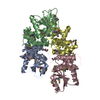


 Mass: 18.015 Da / Num. of mol.: 63 / Source method: isolated from a natural source / Formula: H2O
 Sample preparation
 / Beamline: 19-ID / Wavelength: 0.9792 Å
 Processing