 Movie
Movie Controller

[English] 日本語
 Yorodumi
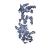
- PDB-2pv7: Crystal structure of chorismate mutase / prephenate dehydrogenase... -
+ Open data

- Basic information
| Entry | Database: PDB / ID: 2pv7 | ||||||
|---|---|---|---|---|---|---|---|
| Title | Crystal structure of chorismate mutase / prephenate dehydrogenase (tyrA) (1574749) from Haemophilus influenzae RD at 2.00 A resolution | ||||||
 Components Components | T-protein [Includes: Chorismate mutase (EC 5.4.99.5) (CM) and Prephenate dehydrogenase (EC 1.3.1.12) (PDH)] | ||||||
 Keywords Keywords |  ISOMERASE / OXIDOREDUCTASE / 1574749 / Chorismate mutase type II / chorismate mutase / prephenate dehydrogenase (tyrA) / Structural Genomics / Joint Center for Structural Genomics / JCSG / Protein Structure Initiative / PSI-2 ISOMERASE / OXIDOREDUCTASE / 1574749 / Chorismate mutase type II / chorismate mutase / prephenate dehydrogenase (tyrA) / Structural Genomics / Joint Center for Structural Genomics / JCSG / Protein Structure Initiative / PSI-2 | ||||||
| Function / homology |  Function and homology informationprephenate dehydrogenase / prephenate dehydrogenase (NADP+) activity / prephenate dehydrogenase (NAD+) activity / tyrosine biosynthetic process / chorismate metabolic process / chorismate mutase / chorismate mutase activity / : / NAD+ binding / cytoplasm Function and homology informationprephenate dehydrogenase / prephenate dehydrogenase (NADP+) activity / prephenate dehydrogenase (NAD+) activity / tyrosine biosynthetic process / chorismate metabolic process / chorismate mutase / chorismate mutase activity / : / NAD+ binding / cytoplasmSimilarity search - Function | ||||||
| Biological species |  Haemophilus influenzae (bacteria) Haemophilus influenzae (bacteria) | ||||||
| Method | X-RAY DIFFRACTION / SYNCHROTRON / MAD / Resolution: 2 Å | ||||||
 Authors Authors | Joint Center for Structural Genomics (JCSG) | ||||||
 Citation Citation | Journal: Acta Crystallogr.,Sect.F / Year: 2010 Title: The structure of Haemophilus influenzae prephenate dehydrogenase suggests unique features of bifunctional TyrA enzymes. Authors: Chiu, H.J. / Abdubek, P. / Astakhova, T. / Axelrod, H.L. / Carlton, D. / Clayton, T. / Das, D. / Deller, M.C. / Duan, L. / Feuerhelm, J. / Grant, J.C. / Grzechnik, A. / Han, G.W. / ...Authors: Chiu, H.J. / Abdubek, P. / Astakhova, T. / Axelrod, H.L. / Carlton, D. / Clayton, T. / Das, D. / Deller, M.C. / Duan, L. / Feuerhelm, J. / Grant, J.C. / Grzechnik, A. / Han, G.W. / Jaroszewski, L. / Jin, K.K. / Klock, H.E. / Knuth, M.W. / Kozbial, P. / Krishna, S.S. / Kumar, A. / Marciano, D. / McMullan, D. / Miller, M.D. / Morse, A.T. / Nigoghossian, E. / Okach, L. / Reyes, R. / Tien, H.J. / Trame, C.B. / van den Bedem, H. / Weekes, D. / Xu, Q. / Hodgson, K.O. / Wooley, J. / Elsliger, M.A. / Deacon, A.M. / Godzik, A. / Lesley, S.A. / Wilson, I.A. | ||||||
| History |
| ||||||
| Remark 300 | BIOMOLECULE: 1 THIS ENTRY CONTAINS THE CRYSTALLOGRAPHIC ASYMMETRIC UNIT WHICH CONSISTS OF 2 CHAINS. ... BIOMOLECULE: 1 THIS ENTRY CONTAINS THE CRYSTALLOGRAPHIC ASYMMETRIC UNIT WHICH CONSISTS OF 2 CHAINS. SEE REMARK 350 FOR INFORMATION ON GENERATING THE BIOLOGICAL MOLECULE(S). SIZE EXCLUSION CHROMATOGRAPHY SUPPORTS THE ASSIGNMENT OF A DIMER AS A SIGNIFICANT OLIGOMERIZATION STATE. | ||||||
| Remark 999 | SEQUENCE THIS CONSTRUCT IS COMPRISED OF AMINO ACIDS 81-377 OF THE FULL-LENGTH PROTEIN (1-377) AND ... SEQUENCE THIS CONSTRUCT IS COMPRISED OF AMINO ACIDS 81-377 OF THE FULL-LENGTH PROTEIN (1-377) AND WAS EXPRESSED WITH A PURIFICATION TAG MGSDKIHHHHHHENLYFQG. THE TAG WAS REMOVED WITH TEV PROTEASE LEAVING ONLY A GLYCINE FOLLOWED BY THE TARGET SEQUENCE. |
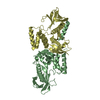
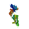
- Structure visualization
| Structure viewer | Molecule: MolmilJmol/JSmol |
|---|
- Downloads & links
-Download
| PDBx/mmCIF format | 2pv7.cif.gz | 133.9 KB | Display | PDBx/mmCIF format |
|---|---|---|---|---|
| PDB format | pdb2pv7.ent.gz | 108.1 KB | Display | PDB format |
| PDBx/mmJSON format | 2pv7.json.gz | Tree view | PDBx/mmJSON format | |
| Others |  Other downloads Other downloads |
-Validation report
| Arichive directory | https://data.pdbj.org/pub/pdb/validation_reports/pv/2pv7ftp://data.pdbj.org/pub/pdb/validation_reports/pv/2pv7 | HTTPS FTP |
|---|
-Related structure data
| Similar structure data | |
|---|---|
| Other databases |

-Links
 PDBj
PDBj


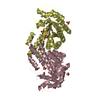
- Assembly
| Deposited unit | 
| |||||||||||||||||||||||||||||||||||||||||||||||||||||||
|---|---|---|---|---|---|---|---|---|---|---|---|---|---|---|---|---|---|---|---|---|---|---|---|---|---|---|---|---|---|---|---|---|---|---|---|---|---|---|---|---|---|---|---|---|---|---|---|---|---|---|---|---|---|---|---|---|
| 1 |
| |||||||||||||||||||||||||||||||||||||||||||||||||||||||
| Unit cell |
| |||||||||||||||||||||||||||||||||||||||||||||||||||||||
| Components on special symmetry positions |
| |||||||||||||||||||||||||||||||||||||||||||||||||||||||
| Noncrystallographic symmetry (NCS) | NCS domain:
NCS domain segments: Ens-ID: 1 / Refine code: 6
| |||||||||||||||||||||||||||||||||||||||||||||||||||||||
| Details | SIZE EXCLUSION CHROMATOGRAPHY WITH STATIC LIGHT SCATTERING SUPPORTS THE ASSIGNMENT OF A DIMER AS THE SIGNIFICANT OLIGOMERIZATION STATE. |
-Components
| #1: Protein | Mass: 34275.805 Da / Num. of mol.: 2 / Fragment: Residues 81-377 Source method: isolated from a genetically manipulated source Source: (gene. exp.) Haemophilus influenzae (bacteria) / Strain: Rd, DSM 11121, KW20 / Gene: 1574749, tyrA, HI1290 / Plasmid: speedET / Production host: Escherichia coli (E. coli) / Strain (production host): HK100References: UniProt: P43902, chorismate mutase, prephenate dehydrogenase#2: Chemical | Tyrosine  Type: L-peptide linking / Mass: 181.189 Da / Num. of mol.: 2 / Source method: obtained synthetically / Formula: C9H11NO3 Type: L-peptide linking / Mass: 181.189 Da / Num. of mol.: 2 / Source method: obtained synthetically / Formula: C9H11NO3#3: Chemical | Nicotinamide adenine dinucleotide  Mass: 663.425 Da / Num. of mol.: 2 / Source method: obtained synthetically / Formula: C21H27N7O14P2 / Comment: NAD*YM Mass: 663.425 Da / Num. of mol.: 2 / Source method: obtained synthetically / Formula: C21H27N7O14P2 / Comment: NAD*YM#4: Water | ChemComp-HOH / | Water Mass: 18.015 Da / Num. of mol.: 393 / Source method: isolated from a natural source / Formula: H2O Mass: 18.015 Da / Num. of mol.: 393 / Source method: isolated from a natural source / Formula: H2O |
|---|
-Experimental details
-Experiment
| Experiment | Method: X-RAY DIFFRACTION / Number of used crystals: 1 |
|---|
- Sample preparation
| Crystal | Density Matthews: 2.98 Å3/Da / Density % sol: 58.72 % |
|---|---|
| Crystal grow | Temperature: 293 K / Method: vapor diffusion, sitting drop Details: NANODROP, 0.04M Potassium dihydrogen phosphate, 20.0% Glycerol, 16.0% PEG 8000, VAPOR DIFFUSION, SITTING DROP, temperature 293K |
-Data collection
| Diffraction | Mean temperature: 100 K | |||||||||||||||||||||||||||||||||||||||||||||||||||||||||||||||||||||||||||||
|---|---|---|---|---|---|---|---|---|---|---|---|---|---|---|---|---|---|---|---|---|---|---|---|---|---|---|---|---|---|---|---|---|---|---|---|---|---|---|---|---|---|---|---|---|---|---|---|---|---|---|---|---|---|---|---|---|---|---|---|---|---|---|---|---|---|---|---|---|---|---|---|---|---|---|---|---|---|---|
| Diffraction source | Source: SYNCHROTRON / Site: SSRL  / Beamline: BL11-1 / Wavelength: 0.91837, 0.97920 / Beamline: BL11-1 / Wavelength: 0.91837, 0.97920 | |||||||||||||||||||||||||||||||||||||||||||||||||||||||||||||||||||||||||||||
| Detector | Type: MARMOSAIC 325 mm CCD / Detector: CCD / Date: Apr 8, 2007 / Details: Flat mirror (vertical focusing) | |||||||||||||||||||||||||||||||||||||||||||||||||||||||||||||||||||||||||||||
| Radiation | Monochromator: Single crystal Si(111) bent (horizontal focusing) Protocol: MAD / Monochromatic (M) / Laue (L): M / Scattering type: x-ray | |||||||||||||||||||||||||||||||||||||||||||||||||||||||||||||||||||||||||||||
| Radiation wavelength |
| |||||||||||||||||||||||||||||||||||||||||||||||||||||||||||||||||||||||||||||
| Reflection | Resolution: 2→29.696 Å / Num. obs: 56593 / % possible obs: 99.7 % / Redundancy: 7.29 % / Biso Wilson estimate: 32.83 Å2 / Rmerge(I) obs: 0.062 / Net I/σ(I): 12.89 | |||||||||||||||||||||||||||||||||||||||||||||||||||||||||||||||||||||||||||||
| Reflection shell |
|
-Phasing
| Phasing | Method: MAD |
|---|
- Processing
| Software |
| |||||||||||||||||||||||||||||||||||||||||||||||||||||||||||||||||||||||||||||||||||||||||||||||||||||||||||||||||||||||||||||||||||||||||||||||||||||||||||||||||||||||||||||||
|---|---|---|---|---|---|---|---|---|---|---|---|---|---|---|---|---|---|---|---|---|---|---|---|---|---|---|---|---|---|---|---|---|---|---|---|---|---|---|---|---|---|---|---|---|---|---|---|---|---|---|---|---|---|---|---|---|---|---|---|---|---|---|---|---|---|---|---|---|---|---|---|---|---|---|---|---|---|---|---|---|---|---|---|---|---|---|---|---|---|---|---|---|---|---|---|---|---|---|---|---|---|---|---|---|---|---|---|---|---|---|---|---|---|---|---|---|---|---|---|---|---|---|---|---|---|---|---|---|---|---|---|---|---|---|---|---|---|---|---|---|---|---|---|---|---|---|---|---|---|---|---|---|---|---|---|---|---|---|---|---|---|---|---|---|---|---|---|---|---|---|---|---|---|---|---|---|
| Refinement | Method to determine structure: MAD / Resolution: 2→29.696 Å / Cor.coef. Fo:Fc: 0.968 / Cor.coef. Fo:Fc free: 0.955 / SU B: 5.931 / SU ML: 0.084 / TLS residual ADP flag: LIKELY RESIDUAL / Cross valid method: THROUGHOUT / σ(F): 0 / ESU R: 0.126 / ESU R Free: 0.121 Stereochemistry target values: MAXIMUM LIKELIHOOD WITH PHASES Details: 1. HYDROGENS HAVE BEEN ADDED IN THE RIDING POSITIONS. 2. A MET-INHIBITION PROTOCOL WAS USED FOR SELENOMETHIONINE INCORPORATION DURING PROTEIN EXPRESSION. THE OCCUPANCY OF THE SE ATOMS IN THE ...Details: 1. HYDROGENS HAVE BEEN ADDED IN THE RIDING POSITIONS. 2. A MET-INHIBITION PROTOCOL WAS USED FOR SELENOMETHIONINE INCORPORATION DURING PROTEIN EXPRESSION. THE OCCUPANCY OF THE SE ATOMS IN THE MSE RESIDUES WAS REDUCED TO 0.75 TO ACCOUNT FOR THE REDUCED SCATTERING POWER DUE TO PARTIAL S-MET INCORPORATION 3. ATOM RECORD CONTAINS RESIDUAL B FACTORS ONLY. 4. RESIDUES 81-91, 312-315, 372-377 IN CHAIN A AND 81-91, 372-377 IN CHAIN B ARE DISORDERED AND ARE NOT MODELED. 5. LIGAND MOLECULES NAD AND TYR ARE MODELED.
| |||||||||||||||||||||||||||||||||||||||||||||||||||||||||||||||||||||||||||||||||||||||||||||||||||||||||||||||||||||||||||||||||||||||||||||||||||||||||||||||||||||||||||||||
| Solvent computation | Ion probe radii: 0.8 Å / Shrinkage radii: 0.8 Å / VDW probe radii: 1.2 Å / Solvent model: MASK | |||||||||||||||||||||||||||||||||||||||||||||||||||||||||||||||||||||||||||||||||||||||||||||||||||||||||||||||||||||||||||||||||||||||||||||||||||||||||||||||||||||||||||||||
| Displacement parameters | Biso mean: 31.26 Å2
| |||||||||||||||||||||||||||||||||||||||||||||||||||||||||||||||||||||||||||||||||||||||||||||||||||||||||||||||||||||||||||||||||||||||||||||||||||||||||||||||||||||||||||||||
| Refinement step | Cycle: LAST / Resolution: 2→29.696 Å
| |||||||||||||||||||||||||||||||||||||||||||||||||||||||||||||||||||||||||||||||||||||||||||||||||||||||||||||||||||||||||||||||||||||||||||||||||||||||||||||||||||||||||||||||
| Refine LS restraints |
| |||||||||||||||||||||||||||||||||||||||||||||||||||||||||||||||||||||||||||||||||||||||||||||||||||||||||||||||||||||||||||||||||||||||||||||||||||||||||||||||||||||||||||||||
| Refine LS restraints NCS | Dom-ID: 1 / Auth asym-ID: A / Ens-ID: 1 / Number: 4218 / Refine-ID: X-RAY DIFFRACTION
| |||||||||||||||||||||||||||||||||||||||||||||||||||||||||||||||||||||||||||||||||||||||||||||||||||||||||||||||||||||||||||||||||||||||||||||||||||||||||||||||||||||||||||||||
| LS refinement shell | Resolution: 2→2.052 Å / Total num. of bins used: 20
| |||||||||||||||||||||||||||||||||||||||||||||||||||||||||||||||||||||||||||||||||||||||||||||||||||||||||||||||||||||||||||||||||||||||||||||||||||||||||||||||||||||||||||||||
| Refinement TLS params. | Method: refined / Refine-ID: X-RAY DIFFRACTION
| |||||||||||||||||||||||||||||||||||||||||||||||||||||||||||||||||||||||||||||||||||||||||||||||||||||||||||||||||||||||||||||||||||||||||||||||||||||||||||||||||||||||||||||||
| Refinement TLS group | Refine-ID: X-RAY DIFFRACTION / Selection: ALL
|
