 Movie
Movie Controller


+ Open data

- Basic information
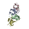
| Entry | Database: PDB / ID: 2pry | ||||||
|---|---|---|---|---|---|---|---|
| Title | Apo form of S. cerevisiae orotate phosphoribosyltransferase | ||||||
 Components Components | Orotate phosphoribosyltransferase 1 | ||||||
 Keywords Keywords | TRANSFERASE / ALPHA BETA / OPRTase apo form | ||||||
| Function / homology |  Function and homology information Function and homology informationpyrimidine ribonucleoside biosynthetic process / orotate phosphoribosyltransferase / orotate phosphoribosyltransferase activity / pyrimidine nucleotide biosynthetic process / 'de novo' UMP biosynthetic process / 'de novo' pyrimidine nucleobase biosynthetic process / nucleus / cytoplasmSimilarity search - Function | ||||||
| Biological species |  Saccharomyces cerevisiae (brewer's yeast) Saccharomyces cerevisiae (brewer's yeast) | ||||||
| Method | X-RAY DIFFRACTION / MOLECULAR REPLACEMENT / Resolution: 2.35 Å | ||||||
 Authors Authors | Gonzalez-Segura, L. / Hurley, T.D. / McClard, R.W. | ||||||
 Citation Citation | Journal: Biochemistry / Year: 2007 Title: Ternary complex formation and induced asymmetry in orotate phosphoribosyltransferase. Authors: Gonzalez-Segura, L. / Witte, J.F. / McClard, R.W. / Hurley, T.D. | ||||||
| History |
|
- Structure visualization
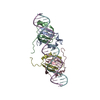
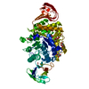
| Structure viewer | Molecule: MolmilJmol/JSmol |
|---|
- Downloads & links
-Download
| PDBx/mmCIF format | 2pry.cif.gz | 57.2 KB | Display | PDBx/mmCIF format |
|---|---|---|---|---|
| PDB format | pdb2pry.ent.gz | 40.3 KB | Display | PDB format |
| PDBx/mmJSON format | 2pry.json.gz | Tree view | PDBx/mmJSON format | |
| Others |  Other downloads Other downloads |
-Validation report
| Arichive directory | https://data.pdbj.org/pub/pdb/validation_reports/pr/2pryftp://data.pdbj.org/pub/pdb/validation_reports/pr/2pry | HTTPS FTP |
|---|
-Related structure data
| Related structure data | 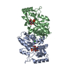 2przC  2ps1C  1stoS C: citing same article ( S: Starting model for refinement |
|---|---|
| Similar structure data |


-Links
 PDBj
PDBj
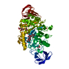
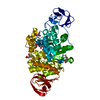
- Assembly
| Deposited unit | 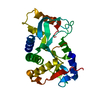
| ||||||||
|---|---|---|---|---|---|---|---|---|---|
| 1 | 
| ||||||||
| Unit cell |
| ||||||||
| Details | The biological assembly is a dimer generated by: -y, -x, 1/2-z |
-Components
| #1: Protein | / OPRT 1 / OPRTase 1 Mass: 24689.439 Da / Num. of mol.: 1 Source method: isolated from a genetically manipulated source Source: (gene. exp.) Saccharomyces cerevisiae (brewer's yeast)Gene: URA5, PYR5 / Plasmid: pREJ2 / Production host:  Escherichia coli (E. coli) / Strain (production host): CS101-4UI Escherichia coli (E. coli) / Strain (production host): CS101-4UIReferences: UniProt: P13298, orotate phosphoribosyltransferase |
|---|---|
| #2: Chemical | ChemComp-MG /   Mass: 24.305 Da / Num. of mol.: 1 / Source method: obtained synthetically / Formula: Mg Mass: 24.305 Da / Num. of mol.: 1 / Source method: obtained synthetically / Formula: Mg |
| #3: Water | ChemComp-HOH / Water Mass: 18.015 Da / Num. of mol.: 100 / Source method: isolated from a natural source / Formula: H2O Mass: 18.015 Da / Num. of mol.: 100 / Source method: isolated from a natural source / Formula: H2O |
-Experimental details
-Experiment
| Experiment | Method: X-RAY DIFFRACTION / Number of used crystals: 1 |
|---|
- Sample preparation
| Crystal | Density Matthews: 2.45 Å3/Da / Density % sol: 49.87 % |
|---|---|
| Crystal grow | Temperature: 293.15 K / Method: vapor diffusion, sitting drop / pH: 7.4 Details: 26% PEG 6000, 0.2M magnesium acetate, 0.1M Tris HCl, pH 7.4, VAPOR DIFFUSION, SITTING DROP, temperature 293.15K |
-Data collection
| Diffraction | Mean temperature: 93.15 K |
|---|---|
| Diffraction source | Source: ROTATING ANODE / Type: RIGAKU RUH2R / Wavelength: 1.54 Å |
| Detector | Type: RIGAKU RAXIS IV / Detector: IMAGE PLATE / Date: Apr 7, 2004 / Details: Osmic confocal |
| Radiation | Monochromator: Osmic confocal / Protocol: SINGLE WAVELENGTH / Monochromatic (M) / Laue (L): M / Scattering type: x-ray |
| Radiation wavelength | Wavelength: 1.54 Å / Relative weight: 1 |
| Reflection | Resolution: 2.35→25 Å / Num. all: 10881 / Num. obs: 10718 / % possible obs: 98.5 % / Observed criterion σ(F): 0 / Observed criterion σ(I): 0.2 / Biso Wilson estimate: 48.52 Å2 / Rmerge(I) obs: 0.053 / Rsym value: 0.053 / Χ2: 1.036 / Net I/σ(I): 24.4 |
| Reflection shell | Resolution: 2.35→2.43 Å / Rmerge(I) obs: 0.246 / Mean I/σ(I) obs: 3.7 / Num. unique all: 870 / Rsym value: 0.246 / Χ2: 1.034 / % possible all: 84.5 |
- Processing
| Software |
| ||||||||||||||||||||||||||||||||||||||||||||||||||||||||||||||||||||||
|---|---|---|---|---|---|---|---|---|---|---|---|---|---|---|---|---|---|---|---|---|---|---|---|---|---|---|---|---|---|---|---|---|---|---|---|---|---|---|---|---|---|---|---|---|---|---|---|---|---|---|---|---|---|---|---|---|---|---|---|---|---|---|---|---|---|---|---|---|---|---|---|
| Refinement | Method to determine structure: MOLECULAR REPLACEMENT Starting model: PDB ENTRY 1STO Resolution: 2.35→24.92 Å / Cor.coef. Fo:Fc: 0.925 / Cor.coef. Fo:Fc free: 0.92 / SU B: 8.323 / SU ML: 0.194 / Cross valid method: THROUGHOUT / σ(F): 0 / σ(I): 0 / ESU R: 0.432 / ESU R Free: 0.26 / Stereochemistry target values: MAXIMUM LIKELIHOOD
| ||||||||||||||||||||||||||||||||||||||||||||||||||||||||||||||||||||||
| Solvent computation | Ion probe radii: 0.8 Å / Shrinkage radii: 0.8 Å / VDW probe radii: 1.4 Å / Solvent model: BABINET MODEL WITH MASK | ||||||||||||||||||||||||||||||||||||||||||||||||||||||||||||||||||||||
| Displacement parameters | Biso mean: 49.601 Å2
| ||||||||||||||||||||||||||||||||||||||||||||||||||||||||||||||||||||||
| Refinement step | Cycle: LAST / Resolution: 2.35→24.92 Å
| ||||||||||||||||||||||||||||||||||||||||||||||||||||||||||||||||||||||
| Refine LS restraints |
| ||||||||||||||||||||||||||||||||||||||||||||||||||||||||||||||||||||||
| LS refinement shell | Resolution: 2.35→2.411 Å / Total num. of bins used: 20
|
