 Movie
Movie Controller

[English] 日本語
 Yorodumi
- PDB-2prs: Structure and metal binding properties of ZnuA, a periplasmic zin... -
+ Open data

- Basic information
| Entry | Database: PDB / ID: 2prs | ||||||
|---|---|---|---|---|---|---|---|
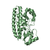
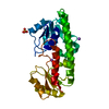
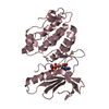
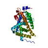
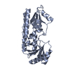
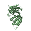
| Title | Structure and metal binding properties of ZnuA, a periplasmic zinc transporter from Escherichia coli | ||||||
 Components Components | High-affinity zinc uptake system protein znuA | ||||||
 Keywords Keywords | METAL TRANSPORT / protein consists of two (beta/alfa)4 domains | ||||||
| Function / homology |  Function and homology information Function and homology informationzinc ion import across plasma membrane / zinc ion transport / ATP-binding cassette (ABC) transporter complex, substrate-binding subunit-containing /  periplasmic space / cell adhesion / zinc ion binding / membrane periplasmic space / cell adhesion / zinc ion binding / membraneSimilarity search - Function | ||||||
| Biological species |  Escherichia coli (E. coli) Escherichia coli (E. coli) | ||||||
| Method | X-RAY DIFFRACTION / SYNCHROTRON / MOLECULAR REPLACEMENT / Resolution: 1.7 Å | ||||||
 Authors Authors | Yatsunyk, L.A. / Kim, L.R. / Vorontsov, I.I. / Rosenzweig, A.C. | ||||||
 Citation Citation | Journal: J.Biol.Inorg.Chem. / Year: 2008 Title: Structure and metal binding properties of ZnuA, a periplasmic zinc transporter from Escherichia coli. Authors: Yatsunyk, L.A. / Easton, J.A. / Kim, L.R. / Sugarbaker, S.A. / Bennett, B. / Breece, R.M. / Vorontsov, I.I. / Tierney, D.L. / Crowder, M.W. / Rosenzweig, A.C. | ||||||
| History |
|
- Structure visualization
| Structure viewer | Molecule: MolmilJmol/JSmol |
|---|
- Downloads & links
-Download
| PDBx/mmCIF format | 2prs.cif.gz | 125.4 KB | Display | PDBx/mmCIF format |
|---|---|---|---|---|
| PDB format | pdb2prs.ent.gz | 96.5 KB | Display | PDB format |
| PDBx/mmJSON format | 2prs.json.gz | Tree view | PDBx/mmJSON format | |
| Others |  Other downloads Other downloads |
-Validation report
| Arichive directory | https://data.pdbj.org/pub/pdb/validation_reports/pr/2prsftp://data.pdbj.org/pub/pdb/validation_reports/pr/2prs | HTTPS FTP |
|---|
-Related structure data


-Links
 PDBj
PDBj
- Assembly
| Deposited unit | 
| ||||||||
|---|---|---|---|---|---|---|---|---|---|
| 1 | 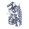
| ||||||||
| 2 | 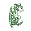
| ||||||||
| Unit cell |
| ||||||||
| Details | the biological assembly is a monomer |
-Components
| #1: Protein | Mass: 31177.219 Da / Num. of mol.: 2 Source method: isolated from a genetically manipulated source Source: (gene. exp.) Escherichia coli (E. coli) / Production host: Escherichia coli (E. coli) / References: UniProt: P39172#2: Chemical |   Mass: 65.409 Da / Num. of mol.: 3 / Source method: obtained synthetically / Formula: Zn Mass: 65.409 Da / Num. of mol.: 3 / Source method: obtained synthetically / Formula: Zn#3: Chemical | ChemComp-IPA / | Isopropyl alcohol  Mass: 60.095 Da / Num. of mol.: 1 / Source method: obtained synthetically / Formula: C3H8O / Comment: alkaloid*YM Mass: 60.095 Da / Num. of mol.: 1 / Source method: obtained synthetically / Formula: C3H8O / Comment: alkaloid*YM#4: Water | ChemComp-HOH / | Water Mass: 18.015 Da / Num. of mol.: 441 / Source method: isolated from a natural source / Formula: H2O Mass: 18.015 Da / Num. of mol.: 441 / Source method: isolated from a natural source / Formula: H2O |
|---|
-Experimental details
-Experiment
| Experiment | Method: X-RAY DIFFRACTION / Number of used crystals: 1 |
|---|
- Sample preparation
| Crystal | Density Matthews: 2.47 Å3/Da / Density % sol: 50.17 % |
|---|---|
| Crystal grow | Temperature: 293 K / Method: vapor diffusion, hanging drop / pH: 7.5 Details: 30 % (w/v) PEG 4000, 5 % isopropanol, 0.1 M HEPES, pH 7.5, VAPOR DIFFUSION, HANGING DROP, temperature 293K |
-Data collection
| Diffraction |
| ||||||||||||||||||||
|---|---|---|---|---|---|---|---|---|---|---|---|---|---|---|---|---|---|---|---|---|---|
| Diffraction source |
| ||||||||||||||||||||
| Detector | Type: MAR scanner 345 mm plate / Detector: IMAGE PLATE / Date: Apr 7, 2006 / Details: mirrors | ||||||||||||||||||||
| Radiation | Monochromator: Si 111 CHANNEL / Protocol: SINGLE WAVELENGTH / Monochromatic (M) / Laue (L): M / Scattering type: x-ray | ||||||||||||||||||||
| Radiation wavelength | Wavelength: 0.97918 Å / Relative weight: 1 | ||||||||||||||||||||
| Reflection | Resolution: 1.7→28.8 Å / Num. obs: 64618 / % possible obs: 97.2 % / Redundancy: 3.6 % / Biso Wilson estimate: 32.5 Å2 / Rsym value: 0.053 | ||||||||||||||||||||
| Reflection shell | Resolution: 1.7→1.76 Å / Redundancy: 3.1 % / Num. unique all: 5643 / Rsym value: 0.323 / % possible all: 85.5 |

- Processing
| Software |
| |||||||||||||||||||||||||||||||||||||||||||||||||||||||||||||||||||||||||||||||||||||||||||||||
|---|---|---|---|---|---|---|---|---|---|---|---|---|---|---|---|---|---|---|---|---|---|---|---|---|---|---|---|---|---|---|---|---|---|---|---|---|---|---|---|---|---|---|---|---|---|---|---|---|---|---|---|---|---|---|---|---|---|---|---|---|---|---|---|---|---|---|---|---|---|---|---|---|---|---|---|---|---|---|---|---|---|---|---|---|---|---|---|---|---|---|---|---|---|---|---|---|
| Refinement | Method to determine structure: MOLECULAR REPLACEMENT Starting model: ZnZnuA in space group P212121 Resolution: 1.7→28.8 Å / Cor.coef. Fo:Fc: 0.954 / Cor.coef. Fo:Fc free: 0.924 / SU B: 2.508 / SU ML: 0.085 / Cross valid method: THROUGHOUT / σ(F): 0 / ESU R: 0.121 / ESU R Free: 0.12 / Stereochemistry target values: MAXIMUM LIKELIHOOD / Details: HYDROGENS HAVE BEEN ADDED IN THE RIDING POSITIONS
| |||||||||||||||||||||||||||||||||||||||||||||||||||||||||||||||||||||||||||||||||||||||||||||||
| Solvent computation | Ion probe radii: 0.8 Å / Shrinkage radii: 0.8 Å / VDW probe radii: 1.2 Å / Solvent model: MASK | |||||||||||||||||||||||||||||||||||||||||||||||||||||||||||||||||||||||||||||||||||||||||||||||
| Displacement parameters | Biso mean: 32.48 Å2
| |||||||||||||||||||||||||||||||||||||||||||||||||||||||||||||||||||||||||||||||||||||||||||||||
| Refinement step | Cycle: LAST / Resolution: 1.7→28.8 Å
| |||||||||||||||||||||||||||||||||||||||||||||||||||||||||||||||||||||||||||||||||||||||||||||||
| Refine LS restraints |
| |||||||||||||||||||||||||||||||||||||||||||||||||||||||||||||||||||||||||||||||||||||||||||||||
| LS refinement shell | Resolution: 1.701→1.745 Å / Total num. of bins used: 20
|
