 Movie
Movie Controller

[English] 日本語
 Yorodumi
- PDB-2pnv: Crystal Structure of the leucine zipper domain of small-conductan... -
+ Open data

- Basic information
| Entry | Database: PDB / ID: 2pnv | ||||||
|---|---|---|---|---|---|---|---|
| Title | Crystal Structure of the leucine zipper domain of small-conductance Ca2+-activated K+ (SKCa) channel from Rattus norvegicus | ||||||
 Components Components | Small conductance calcium-activated potassium channel protein 2 | ||||||
 Keywords Keywords |  MEMBRANE PROTEIN / leucine zipper / SKCa channel / Rattus norvegicus MEMBRANE PROTEIN / leucine zipper / SKCa channel / Rattus norvegicus | ||||||
| Function / homology |  Function and homology information Function and homology informationCa2+ activated K+ channels / small conductance calcium-activated potassium channel activity / membrane repolarization during atrial cardiac muscle cell action potential / calcium-activated potassium channel activity / positive regulation of potassium ion transport / inward rectifier potassium channel activity / regulation of potassium ion transmembrane transport / regulation of neuronal synaptic plasticity / alpha-actinin binding / smooth endoplasmic reticulum ...Ca2+ activated K+ channels / small conductance calcium-activated potassium channel activity / membrane repolarization during atrial cardiac muscle cell action potential / calcium-activated potassium channel activity / positive regulation of potassium ion transport / inward rectifier potassium channel activity / regulation of potassium ion transmembrane transport / regulation of neuronal synaptic plasticity / alpha-actinin binding / smooth endoplasmic reticulum / potassium ion transmembrane transport / T-tubule / modulation of chemical synaptic transmission / potassium ion transport / sarcolemma / Z disc / postsynaptic membrane / dendritic spine / calmodulin binding / protein domain specific binding / neuronal cell body / glutamatergic synapse / cell surface / protein homodimerization activity / membrane / plasma membraneSimilarity search - Function | ||||||
| Biological species |  Rattus norvegicus (Norway rat) Rattus norvegicus (Norway rat) | ||||||
| Method | X-RAY DIFFRACTION / SYNCHROTRON / MOLECULAR REPLACEMENT / Resolution: 2.1 Å | ||||||
 Authors Authors | Kim, J.Y. / Kim, M.K. / Kang, G.B. / Park, C.S. / Eom, S.H. | ||||||
 Citation Citation | Journal: Proteins / Year: 2008 Title: Crystal structure of the leucine zipper domain of small-conductance Ca2+-activated K+ (SK(Ca)) channel from Rattus norvegicus. Authors: Kim, J.Y. / Kim, M.K. / Kang, G.B. / Park, C.S. / Eom, S.H. | ||||||
| History |
|
- Structure visualization
| Structure viewer | Molecule: MolmilJmol/JSmol |
|---|
- Downloads & links
-Download
| PDBx/mmCIF format | 2pnv.cif.gz | 27.5 KB | Display | PDBx/mmCIF format |
|---|---|---|---|---|
| PDB format | pdb2pnv.ent.gz | 18.2 KB | Display | PDB format |
| PDBx/mmJSON format | 2pnv.json.gz | Tree view | PDBx/mmJSON format | |
| Others |  Other downloads Other downloads |
-Validation report
| Arichive directory | https://data.pdbj.org/pub/pdb/validation_reports/pn/2pnvftp://data.pdbj.org/pub/pdb/validation_reports/pn/2pnv | HTTPS FTP |
|---|
-Related structure data
| Related structure data |  1c94S S: Starting model for refinement |
|---|---|
| Similar structure data |
-Links
 PDBj
PDBj

- Assembly
| Deposited unit | 
| ||||||||
|---|---|---|---|---|---|---|---|---|---|
| 1 | 
| ||||||||
| 2 | 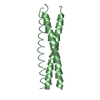
| ||||||||
| 3 | 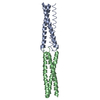
| ||||||||
| Unit cell |
|
-Components
| #1: Protein/peptide | Mass: 4954.655 Da / Num. of mol.: 2 / Fragment: leucine zipper domain Source method: isolated from a genetically manipulated source Source: (gene. exp.) Rattus norvegicus (Norway rat) / Gene: Kcnn2 / Plasmid: modified pGEX vector / Species (production host): Escherichia coli / Production host:  Escherichia coli BL21(DE3) (bacteria) / Strain (production host): BL21(DE3) / References: UniProt: P70604 Escherichia coli BL21(DE3) (bacteria) / Strain (production host): BL21(DE3) / References: UniProt: P70604#2: Water | ChemComp-HOH / | Water Mass: 18.015 Da / Num. of mol.: 4 / Source method: isolated from a natural source / Formula: H2O Mass: 18.015 Da / Num. of mol.: 4 / Source method: isolated from a natural source / Formula: H2O |
|---|
-Experimental details
-Experiment
| Experiment | Method: X-RAY DIFFRACTION / Number of used crystals: 1 |
|---|
- Sample preparation
| Crystal | Density Matthews: 2.257 Å3/Da / Density % sol: 43.41 % |
|---|---|
| Crystal grow | Temperature: 298 K / Method: vapor diffusion, hanging drop / pH: 5.6 Details: 0.1M sodium citrate (pH 5.6), 20-22% (w/v) PEG 8000, VAPOR DIFFUSION, HANGING DROP, temperature 298.0K |
-Data collection
| Diffraction | Mean temperature: 200 K |
|---|---|
| Diffraction source | Source: SYNCHROTRON / Site: Photon Factory  / Beamline: BL-5A / Wavelength: 1 Å / Beamline: BL-5A / Wavelength: 1 Å |
| Detector | Detector: CCD / Date: Nov 7, 2004 |
| Radiation | Protocol: SINGLE WAVELENGTH / Monochromatic (M) / Laue (L): M / Scattering type: x-ray |
| Radiation wavelength | Wavelength: 1 Å / Relative weight: 1 |
| Reflection twin | Type: hemihedral / Operator: h,-h-k,-l / Fraction: 0.35 |
| Reflection | Resolution: 2.1→50 Å / Num. all: 5216 / Num. obs: 5204 / % possible obs: 99.8 % / Observed criterion σ(F): 0 / Observed criterion σ(I): 0 / Redundancy: 15.8 % / Rmerge(I) obs: 0.062 / Rsym value: 0.062 / Net I/σ(I): 68.1 |
| Reflection shell | Resolution: 2.1→2.14 Å / Redundancy: 14.8 % / Rmerge(I) obs: 0.324 / Mean I/σ(I) obs: 8.2 / Num. unique all: 232 / Rsym value: 0.324 / % possible all: 99.6 |
- Processing
| Software |
| |||||||||||||||||||||||||
|---|---|---|---|---|---|---|---|---|---|---|---|---|---|---|---|---|---|---|---|---|---|---|---|---|---|---|
| Refinement | Method to determine structure: MOLECULAR REPLACEMENT Starting model: PDB entry 1C94 Resolution: 2.1→20 Å / Isotropic thermal model: anisotropic / Cross valid method: THROUGHOUT / σ(F): 0 / σ(I): 0 / Stereochemistry target values: Engh & Huber Details: THIS IS A TWINNED STRUCTURE. THE TWINNING OPERATOR IS (H,K,L) -> (H,-H-K,-L) AND THE TWINNING FRACTION IS 0.35. THE R-FACTOR IS 0.210 AND THE R-FREE IS 0.278 WHEN THIS TWINNING OPERATOR IS USED.
| |||||||||||||||||||||||||
| Displacement parameters | Biso mean: 52.3 Å2
| |||||||||||||||||||||||||
| Refine analyze | Luzzati coordinate error obs: 0.37 Å / Luzzati d res low obs: 5 Å / Luzzati sigma a obs: 0.36 Å | |||||||||||||||||||||||||
| Refinement step | Cycle: LAST / Resolution: 2.1→20 Å
| |||||||||||||||||||||||||
| Refine LS restraints |
| |||||||||||||||||||||||||
| LS refinement shell | Resolution: 2.1→2.23 Å / Rfactor Rfree error: 0.046
|
