 Movie
Movie Controller


+ Open data

- Basic information
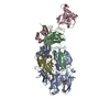
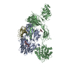
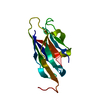
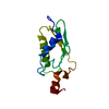
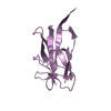
| Entry | Database: PDB / ID: 2pnd | ||||||
|---|---|---|---|---|---|---|---|
| Title | Structure or murine CRIg | ||||||
 Components Components | V-set and immunoglobulin domain containing 4 | ||||||
 Keywords Keywords | IMMUNE SYSTEM / Complement Receptor / Ig-Like domain | ||||||
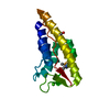
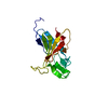
| Function / homology | Immunoglobulins / Immunoglobulin-like / Sandwich / Mainly Beta / :  Function and homology information Function and homology information | ||||||
| Biological species |  Mus musculus (house mouse) Mus musculus (house mouse) | ||||||
| Method | X-RAY DIFFRACTION / SYNCHROTRON / MOLECULAR REPLACEMENT / Resolution: 1 Å | ||||||
 Authors Authors | Wiesmann, C. | ||||||
 Citation Citation | Journal: J.Exp.Med. / Year: 2007 Title: A novel inhibitor of the alternative pathway of complement reverses inflammation and bone destruction in experimental arthritis. Authors: Katschke, K.J. / Helmy, K.Y. / Steffek, M. / Xi, H. / Yin, J. / Lee, W.P. / Gribling, P. / Barck, K.H. / Carano, R.A. / Taylor, R.E. / Rangell, L. / Diehl, L. / Hass, P.E. / Wiesmann, C. / ...Authors: Katschke, K.J. / Helmy, K.Y. / Steffek, M. / Xi, H. / Yin, J. / Lee, W.P. / Gribling, P. / Barck, K.H. / Carano, R.A. / Taylor, R.E. / Rangell, L. / Diehl, L. / Hass, P.E. / Wiesmann, C. / van Lookerenb Campagne, M. | ||||||
| History |
|
- Structure visualization
| Structure viewer | Molecule: MolmilJmol/JSmol |
|---|
- Downloads & links
-Download
| PDBx/mmCIF format | 2pnd.cif.gz | 71.4 KB | Display | PDBx/mmCIF format |
|---|---|---|---|---|
| PDB format | pdb2pnd.ent.gz | 52.4 KB | Display | PDB format |
| PDBx/mmJSON format | 2pnd.json.gz | Tree view | PDBx/mmJSON format | |
| Others |  Other downloads Other downloads |
-Validation report
| Arichive directory | https://data.pdbj.org/pub/pdb/validation_reports/pn/2pndftp://data.pdbj.org/pub/pdb/validation_reports/pn/2pnd | HTTPS FTP |
|---|
-Related structure data
| Related structure data |  2iccS S: Starting model for refinement |
|---|---|
| Similar structure data |


-Links
 PDBj
PDBj- Assembly
| Deposited unit | 
| ||||||||
|---|---|---|---|---|---|---|---|---|---|
| 1 |
| ||||||||
| Unit cell |
|
-Components
| #1: Protein | Mass: 13761.718 Da / Num. of mol.: 1 Source method: isolated from a genetically manipulated source Source: (gene. exp.) Mus musculus (house mouse) / Gene: Vsig4, BC025105 / Production host:  Escherichia coli (E. coli) / References: UniProt: Q80WA3 Escherichia coli (E. coli) / References: UniProt: Q80WA3 |
|---|---|
| #2: Water | ChemComp-HOH / Water Mass: 18.015 Da / Num. of mol.: 237 / Source method: isolated from a natural source / Formula: H2O Mass: 18.015 Da / Num. of mol.: 237 / Source method: isolated from a natural source / Formula: H2O |
-Experimental details
-Experiment
| Experiment | Method: X-RAY DIFFRACTION / Number of used crystals: 1 |
|---|
- Sample preparation
| Crystal | Density Matthews: 1.81 Å3/Da / Density % sol: 32.1 % |
|---|---|
| Crystal grow | Temperature: 298 K / Method: evaporation / pH: 7.5 Details: 100 mM NaCl, 25 mM TRIS, 20 % PEG 3350, 0.2 M Potassium tartrate, pH 7.5, EVAPORATION, temperature 298K |
-Data collection
| Diffraction | Mean temperature: 100 K |
|---|---|
| Diffraction source | Source: SYNCHROTRON / Site: ALS  / Beamline: 5.0.2 / Wavelength: 1 Å / Beamline: 5.0.2 / Wavelength: 1 Å |
| Detector | Type: ADSC QUANTUM 315 / Detector: CCD / Date: Feb 6, 2005 |
| Radiation | Protocol: SINGLE WAVELENGTH / Monochromatic (M) / Laue (L): M / Scattering type: x-ray |
| Radiation wavelength | Wavelength: 1 Å / Relative weight: 1 |
| Reflection | Resolution: 0.97→50 Å / Num. all: 60032 / Num. obs: 51825 / % possible obs: 86.3 % / Observed criterion σ(F): 0 / Observed criterion σ(I): 0 / Redundancy: 6.2 % / Rsym value: 0.41 / Net I/σ(I): 15.7 |
| Reflection shell | Resolution: 0.97→1 Å / Redundancy: 1.6 % / Mean I/σ(I) obs: 2.7 / Rsym value: 0.21 / % possible all: 24.9 |
- Processing
| Software |
| ||||||||||||||||||||||||||||||||||||||||||||||||||||||||||||||||||||||||||||||||||||||||||||||||||||||||||||||||||||||||||||||||||||||||||||||||||||||||||||||||||||||||||
|---|---|---|---|---|---|---|---|---|---|---|---|---|---|---|---|---|---|---|---|---|---|---|---|---|---|---|---|---|---|---|---|---|---|---|---|---|---|---|---|---|---|---|---|---|---|---|---|---|---|---|---|---|---|---|---|---|---|---|---|---|---|---|---|---|---|---|---|---|---|---|---|---|---|---|---|---|---|---|---|---|---|---|---|---|---|---|---|---|---|---|---|---|---|---|---|---|---|---|---|---|---|---|---|---|---|---|---|---|---|---|---|---|---|---|---|---|---|---|---|---|---|---|---|---|---|---|---|---|---|---|---|---|---|---|---|---|---|---|---|---|---|---|---|---|---|---|---|---|---|---|---|---|---|---|---|---|---|---|---|---|---|---|---|---|---|---|---|---|---|---|---|
| Refinement | Method to determine structure: MOLECULAR REPLACEMENT Starting model: 2ICC Resolution: 1→39.53 Å / Cor.coef. Fo:Fc: 0.978 / Cor.coef. Fo:Fc free: 0.973 / SU B: 0.553 / SU ML: 0.014 / Cross valid method: THROUGHOUT / ESU R: 0.023 / ESU R Free: 0.023 / Stereochemistry target values: MAXIMUM LIKELIHOOD / Details: HYDROGENS HAVE BEEN ADDED IN THE RIDING POSITIONS
| ||||||||||||||||||||||||||||||||||||||||||||||||||||||||||||||||||||||||||||||||||||||||||||||||||||||||||||||||||||||||||||||||||||||||||||||||||||||||||||||||||||||||||
| Solvent computation | Ion probe radii: 0.8 Å / Shrinkage radii: 0.8 Å / VDW probe radii: 1.4 Å / Solvent model: BABINET MODEL WITH MASK | ||||||||||||||||||||||||||||||||||||||||||||||||||||||||||||||||||||||||||||||||||||||||||||||||||||||||||||||||||||||||||||||||||||||||||||||||||||||||||||||||||||||||||
| Displacement parameters | Biso mean: 8.979 Å2
| ||||||||||||||||||||||||||||||||||||||||||||||||||||||||||||||||||||||||||||||||||||||||||||||||||||||||||||||||||||||||||||||||||||||||||||||||||||||||||||||||||||||||||
| Refinement step | Cycle: LAST / Resolution: 1→39.53 Å
| ||||||||||||||||||||||||||||||||||||||||||||||||||||||||||||||||||||||||||||||||||||||||||||||||||||||||||||||||||||||||||||||||||||||||||||||||||||||||||||||||||||||||||
| Refine LS restraints |
| ||||||||||||||||||||||||||||||||||||||||||||||||||||||||||||||||||||||||||||||||||||||||||||||||||||||||||||||||||||||||||||||||||||||||||||||||||||||||||||||||||||||||||
| LS refinement shell | Resolution: 1→1.021 Å / Total num. of bins used: 25
| ||||||||||||||||||||||||||||||||||||||||||||||||||||||||||||||||||||||||||||||||||||||||||||||||||||||||||||||||||||||||||||||||||||||||||||||||||||||||||||||||||||||||||
| Refinement TLS params. | Method: refined / Origin x: 9.0292 Å / Origin y: 28.7997 Å / Origin z: 9.376 Å
|
