 Movie
Movie Controller


+ Open data

- Basic information
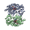
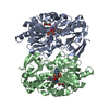
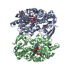
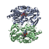
| Entry | Database: PDB / ID: 2pml | ||||||
|---|---|---|---|---|---|---|---|
| Title | Crystal structure of PfPK7 in complex with an ATP analogue | ||||||
 Components Components | Ser/Thr protein kinase Serine/threonine-specific protein kinase Serine/threonine-specific protein kinase | ||||||
 Keywords Keywords | TRANSFERASE / Ser/Thr protein kinase / Plasmodium falciparum / phosphorylation | ||||||
| Function / homology |  Function and homology information Function and homology informationJUN kinase kinase activity / mitotic DNA damage checkpoint signaling / mitogen-activated protein kinase kinase / MAP kinase kinase activity / protein kinase activity / phosphorylation / protein serine/threonine kinase activity / ATP binding / nucleus / cytoplasmSimilarity search - Function | ||||||
| Biological species |  Plasmodium falciparum (malaria parasite P. falciparum) Plasmodium falciparum (malaria parasite P. falciparum) | ||||||
| Method | X-RAY DIFFRACTION / SYNCHROTRON / MAD / Resolution: 2.6 Å | ||||||
 Authors Authors | Merckx, A. / Echalier, A. / Noble, M. / Endicott, J. | ||||||
 Citation Citation | Journal: To be Published Title: Structures of PfPK7 an atypical protein kinase from P. falciparum identify a novel activation motif and leads for inhibitor design Authors: Merckx, A. / Echalier, A. / Langford, K. / Sicard, A. / Langsley, G. / Joore, J. / Doerig, C. / Noble, M. / Endicott, J. | ||||||
| History |
| ||||||
| Remark 300 | THIS ENTRY CONTAINS THE CRYSTALLOGRAPHIC ASYMMETRIC UNIT WHICH CONSISTS OF 1 CHAIN(S). Authors ... THIS ENTRY CONTAINS THE CRYSTALLOGRAPHIC ASYMMETRIC UNIT WHICH CONSISTS OF 1 CHAIN(S). Authors state that the biological unit is unknown. |
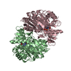
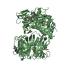
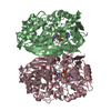
- Structure visualization
| Structure viewer | Molecule: MolmilJmol/JSmol |
|---|
- Downloads & links
-Download
| PDBx/mmCIF format | 2pml.cif.gz | 86 KB | Display | PDBx/mmCIF format |
|---|---|---|---|---|
| PDB format | pdb2pml.ent.gz | 63.9 KB | Display | PDB format |
| PDBx/mmJSON format | 2pml.json.gz | Tree view | PDBx/mmJSON format | |
| Others |  Other downloads Other downloads |
-Validation report
| Arichive directory | https://data.pdbj.org/pub/pdb/validation_reports/pm/2pmlftp://data.pdbj.org/pub/pdb/validation_reports/pm/2pml | HTTPS FTP |
|---|
-Related structure data
| Similar structure data |
|---|


-Links
 PDBj
PDBj


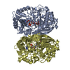
- Assembly
| Deposited unit | 
| ||||||||
|---|---|---|---|---|---|---|---|---|---|
| 1 | 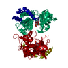
| ||||||||
| Unit cell |
| ||||||||
| Details | AUTHORS STATE THAT THE BIOLOGICAL UNIT IS UNKNOWN |
-Components
| #1: Protein | Serine/threonine-specific protein kinase / PfPK7 Mass: 41434.598 Da / Num. of mol.: 1 Source method: isolated from a genetically manipulated source Source: (gene. exp.) Plasmodium falciparum (malaria parasite P. falciparum)Gene: PFB0605w / Plasmid: pGEX / Species (production host): Escherichia coli / Production host:  Escherichia coli BL21 (bacteria) / Strain (production host): BL21 / References: UniProt: Q7YTF7, UniProt: O96214*PLUS Escherichia coli BL21 (bacteria) / Strain (production host): BL21 / References: UniProt: Q7YTF7, UniProt: O96214*PLUS |
|---|---|
| #2: Chemical | ChemComp-MN /   Mass: 54.938 Da / Num. of mol.: 1 / Source method: obtained synthetically / Formula: Mn Mass: 54.938 Da / Num. of mol.: 1 / Source method: obtained synthetically / Formula: Mn |
| #3: Chemical | ChemComp-ANP /   Mass: 506.196 Da / Num. of mol.: 1 / Source method: obtained synthetically / Formula: C10H17N6O12P3 / Comment: AMP-PNP, energy-carrying molecule analogue*YM Mass: 506.196 Da / Num. of mol.: 1 / Source method: obtained synthetically / Formula: C10H17N6O12P3 / Comment: AMP-PNP, energy-carrying molecule analogue*YM |
| #4: Water | ChemComp-HOH / Water Mass: 18.015 Da / Num. of mol.: 27 / Source method: isolated from a natural source / Formula: H2O Mass: 18.015 Da / Num. of mol.: 27 / Source method: isolated from a natural source / Formula: H2O |
-Experimental details
-Experiment
| Experiment | Method: X-RAY DIFFRACTION / Number of used crystals: 1 |
|---|
- Sample preparation
| Crystal | Density Matthews: 2.47 Å3/Da / Density % sol: 50.3 % |
|---|---|
| Crystal grow | Temperature: 300 K / Method: vapor diffusion, sitting drop / pH: 7.5 Details: 0.1 M Hepes pH7.5; 20% PEG 10K, VAPOR DIFFUSION, SITTING DROP, temperature 300K |
-Data collection
| Diffraction | Mean temperature: 100 K | |||||||||||||||
|---|---|---|---|---|---|---|---|---|---|---|---|---|---|---|---|---|
| Diffraction source | Source: SYNCHROTRON / Site: ESRF  / Beamline: BM14 / Wavelength: 0.9762, 0.9791, 0.9792, 0.9686 / Beamline: BM14 / Wavelength: 0.9762, 0.9791, 0.9792, 0.9686 | |||||||||||||||
| Detector | Type: MARMOSAIC 225 mm CCD / Detector: CCD / Date: May 10, 2005 | |||||||||||||||
| Radiation | Monochromator: Si 111 / Protocol: MAD / Monochromatic (M) / Laue (L): M / Scattering type: x-ray | |||||||||||||||
| Radiation wavelength |
| |||||||||||||||
| Reflection | Resolution: 2.6→20 Å / Num. obs: 13019 / % possible obs: 100 % / Observed criterion σ(F): 2 / Observed criterion σ(I): 2 / Redundancy: 6.7 % / Rmerge(I) obs: 0.05 / Net I/σ(I): 24.1 | |||||||||||||||
| Reflection shell | Resolution: 2.6→2.666 Å / Rmerge(I) obs: 0.424 / Mean I/σ(I) obs: 4.9 / % possible all: 100 |
- Processing
| Software |
| ||||||||||||||||||||||||||||||||||||||||||||||||||||||||||||||||||||||||||||||||||||||||||
|---|---|---|---|---|---|---|---|---|---|---|---|---|---|---|---|---|---|---|---|---|---|---|---|---|---|---|---|---|---|---|---|---|---|---|---|---|---|---|---|---|---|---|---|---|---|---|---|---|---|---|---|---|---|---|---|---|---|---|---|---|---|---|---|---|---|---|---|---|---|---|---|---|---|---|---|---|---|---|---|---|---|---|---|---|---|---|---|---|---|---|---|
| Refinement | Method to determine structure: MAD / Resolution: 2.6→20 Å / Cor.coef. Fo:Fc: 0.913 / Cor.coef. Fo:Fc free: 0.884 / SU B: 30.868 / SU ML: 0.346 / TLS residual ADP flag: LIKELY RESIDUAL / Cross valid method: THROUGHOUT / σ(F): 2 / ESU R: 1.477 / ESU R Free: 0.421 / Stereochemistry target values: MAXIMUM LIKELIHOOD
| ||||||||||||||||||||||||||||||||||||||||||||||||||||||||||||||||||||||||||||||||||||||||||
| Solvent computation | Ion probe radii: 0.8 Å / Shrinkage radii: 0.8 Å / VDW probe radii: 1.4 Å / Solvent model: MASK | ||||||||||||||||||||||||||||||||||||||||||||||||||||||||||||||||||||||||||||||||||||||||||
| Displacement parameters | Biso mean: 43.535 Å2
| ||||||||||||||||||||||||||||||||||||||||||||||||||||||||||||||||||||||||||||||||||||||||||
| Refinement step | Cycle: LAST / Resolution: 2.6→20 Å
| ||||||||||||||||||||||||||||||||||||||||||||||||||||||||||||||||||||||||||||||||||||||||||
| Refine LS restraints |
| ||||||||||||||||||||||||||||||||||||||||||||||||||||||||||||||||||||||||||||||||||||||||||
| LS refinement shell | Resolution: 2.6→2.666 Å / Total num. of bins used: 20
| ||||||||||||||||||||||||||||||||||||||||||||||||||||||||||||||||||||||||||||||||||||||||||
| Refinement TLS params. | Method: refined / Origin x: -12.052 Å / Origin y: 16.63 Å / Origin z: -20.748 Å
|
