 Movie
Movie Controller

[English] 日本語
 Yorodumi
- PDB-2pk2: Cyclin box structure of the P-TEFb subunit Cyclin T1 derived from... -
+ Open data

- Basic information
| Entry | Database: PDB / ID: 2pk2 | ||||||
|---|---|---|---|---|---|---|---|
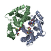
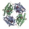
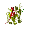
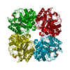
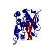
| Title | Cyclin box structure of the P-TEFb subunit Cyclin T1 derived from a fusion complex with EIAV Tat | ||||||
 Components Components | Cyclin-T1, Protein Tat | ||||||
 Keywords Keywords |  CELL CYCLE / CYCLIN T1 / TAT / TAR / TWINNING / TRANSCRIPTION REGULATION P-TEFb CELL CYCLE / CYCLIN T1 / TAT / TAR / TWINNING / TRANSCRIPTION REGULATION P-TEFb | ||||||
| Function / homology |  Function and homology informationP-TEFb complex / Interactions of Tat with host cellular proteins / 7SK snRNA binding / positive regulation of viral transcription / cyclin/CDK positive transcription elongation factor complex / host cell nucleolus / cyclin-dependent protein serine/threonine kinase activator activity / positive regulation by host of viral transcription / RNA polymerase binding / positive regulation of DNA-templated transcription, elongation ...P-TEFb complex / Interactions of Tat with host cellular proteins / 7SK snRNA binding / positive regulation of viral transcription / cyclin/CDK positive transcription elongation factor complex / host cell nucleolus / cyclin-dependent protein serine/threonine kinase activator activity / positive regulation by host of viral transcription / RNA polymerase binding / positive regulation of DNA-templated transcription, elongation / regulation of cyclin-dependent protein serine/threonine kinase activity / Pausing and recovery of Tat-mediated HIV elongation / Tat-mediated HIV elongation arrest and recovery / HIV elongation arrest and recovery / Pausing and recovery of HIV elongation / RNA polymerase II transcribes snRNA genes / RNA-binding transcription regulator activity / Tat-mediated elongation of the HIV-1 transcript / Formation of HIV-1 elongation complex containing HIV-1 Tat / Formation of HIV elongation complex in the absence of HIV Tat / RNA Polymerase II Transcription Elongation / Formation of RNA Pol II elongation complex / RNA Polymerase II Pre-transcription Events / viral process / molecular condensate scaffold activity / TP53 Regulates Transcription of DNA Repair Genes / positive regulation of transcription elongation by RNA polymerase II / SMAD2/SMAD3:SMAD4 heterotrimer regulates transcription / DNA-binding transcription factor binding / Estrogen-dependent gene expression / transcription by RNA polymerase II / transcription cis-regulatory region binding / response to xenobiotic stimulus / cell cycle / cell division / chromatin binding / regulation of transcription by RNA polymerase II / protein kinase binding / positive regulation of transcription by RNA polymerase II / DNA binding / RNA binding / nucleoplasm / nucleus / cytosol Function and homology informationP-TEFb complex / Interactions of Tat with host cellular proteins / 7SK snRNA binding / positive regulation of viral transcription / cyclin/CDK positive transcription elongation factor complex / host cell nucleolus / cyclin-dependent protein serine/threonine kinase activator activity / positive regulation by host of viral transcription / RNA polymerase binding / positive regulation of DNA-templated transcription, elongation ...P-TEFb complex / Interactions of Tat with host cellular proteins / 7SK snRNA binding / positive regulation of viral transcription / cyclin/CDK positive transcription elongation factor complex / host cell nucleolus / cyclin-dependent protein serine/threonine kinase activator activity / positive regulation by host of viral transcription / RNA polymerase binding / positive regulation of DNA-templated transcription, elongation / regulation of cyclin-dependent protein serine/threonine kinase activity / Pausing and recovery of Tat-mediated HIV elongation / Tat-mediated HIV elongation arrest and recovery / HIV elongation arrest and recovery / Pausing and recovery of HIV elongation / RNA polymerase II transcribes snRNA genes / RNA-binding transcription regulator activity / Tat-mediated elongation of the HIV-1 transcript / Formation of HIV-1 elongation complex containing HIV-1 Tat / Formation of HIV elongation complex in the absence of HIV Tat / RNA Polymerase II Transcription Elongation / Formation of RNA Pol II elongation complex / RNA Polymerase II Pre-transcription Events / viral process / molecular condensate scaffold activity / TP53 Regulates Transcription of DNA Repair Genes / positive regulation of transcription elongation by RNA polymerase II / SMAD2/SMAD3:SMAD4 heterotrimer regulates transcription / DNA-binding transcription factor binding / Estrogen-dependent gene expression / transcription by RNA polymerase II / transcription cis-regulatory region binding / response to xenobiotic stimulus / cell cycle / cell division / chromatin binding / regulation of transcription by RNA polymerase II / protein kinase binding / positive regulation of transcription by RNA polymerase II / DNA binding / RNA binding / nucleoplasm / nucleus / cytosolSimilarity search - Function | ||||||
| Biological species |  Homo sapiens (human) Homo sapiens (human) Equine infectious anemia virus Equine infectious anemia virus | ||||||
| Method | X-RAY DIFFRACTION / SYNCHROTRON / MOLECULAR REPLACEMENT / Resolution: 2.67 Å | ||||||
 Authors Authors | Anand, K. / Schulte, A. / Fujinaga, K. / Scheffzek, K. / Geyer, M. | ||||||
 Citation Citation | Journal: J.Mol.Biol. / Year: 2007 Title: Cyclin Box Structure of the P-TEFb Subunit Cyclin T1 Derived from a Fusion Complex with EIAV Tat. Authors: Anand, K. / Schulte, A. / Fujinaga, K. / Scheffzek, K. / Geyer, M. | ||||||
| History |
| ||||||
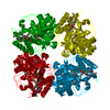
| Remark 300 | BIOMOLECULE: 1 THIS ENTRY CONTAINS THE CRYSTALLOGRAPHIC ASYMMETRIC UNIT WHICH CONSISTS OF 4 CHAIN(S) ...BIOMOLECULE: 1 THIS ENTRY CONTAINS THE CRYSTALLOGRAPHIC ASYMMETRIC UNIT WHICH CONSISTS OF 4 CHAIN(S). THE BIOLOGICAL ASSEMBLY IS A TETRAMER, GENERATED FROM TWO DIMERS IN THE ASYMMETRIC UNIT BY TWIN OPERATIONS:1/3 2/3 2/3, -1/3 1/3 -2/3, -4/3 -2/3 1/3, 4. SEE REMARK 350 FOR INFORMATION ON GENERATING THE BIOLOGICAL MOLECULE(S). | ||||||
| Remark 999 | SEQUENCE Authors state the nucleotide sequence is GenBank entry AF048730 (human Cyclin T1), which ...SEQUENCE Authors state the nucleotide sequence is GenBank entry AF048730 (human Cyclin T1), which refers to the GenPept entry AAC39664. Residue 77 is Gln in UNP entry O60563, but is Arg in AF048730. Authors state the electron density map accommodates Arg side chain very well. |
- Structure visualization
| Structure viewer | Molecule: MolmilJmol/JSmol |
|---|
- Downloads & links
-Download
| PDBx/mmCIF format | 2pk2.cif.gz | 214.1 KB | Display | PDBx/mmCIF format |
|---|---|---|---|---|
| PDB format | pdb2pk2.ent.gz | 171.4 KB | Display | PDB format |
| PDBx/mmJSON format | 2pk2.json.gz | Tree view | PDBx/mmJSON format | |
| Others |  Other downloads Other downloads |
-Validation report
| Arichive directory | https://data.pdbj.org/pub/pdb/validation_reports/pk/2pk2ftp://data.pdbj.org/pub/pdb/validation_reports/pk/2pk2 | HTTPS FTP |
|---|
-Related structure data
| Related structure data | |
|---|---|
| Similar structure data |


-Links
 PDBj
PDBj


- Assembly
| Deposited unit | 
| ||||||||
|---|---|---|---|---|---|---|---|---|---|
| 1 |
| ||||||||
| Unit cell |
| ||||||||
| Details | The biological assembly is a tetramer, generated from two dimers in the asymmetric unit by twin operations:1/3 2/3 2/3, -1/3 1/3 -2/3, -4/3 -2/3 1/3, 4 |
-Components
| #1: Protein | Mass: 40341.637 Da / Num. of mol.: 4 Fragment: fusion protein of Cyclin T1 residues 1-281 and Protein EIAV Tat residues 19-75 Source method: isolated from a genetically manipulated source Source: (gene. exp.) Homo sapiens (human), (gene. exp.) Equine infectious anemia virusGenus: Homo, Lentivirus / Species: , / Strain: , / Plasmid: pGEX-4T1 / Species (production host): Escherichia coli / Production host:  Escherichia coli BL21(DE3) (bacteria) / Strain (production host): BL21(DE3) / References: UniProt: O60563, UniProt: P32544 Escherichia coli BL21(DE3) (bacteria) / Strain (production host): BL21(DE3) / References: UniProt: O60563, UniProt: P32544#2: Water | ChemComp-HOH / | Water Mass: 18.015 Da / Num. of mol.: 79 / Source method: isolated from a natural source / Formula: H2O Mass: 18.015 Da / Num. of mol.: 79 / Source method: isolated from a natural source / Formula: H2O |
|---|
-Experimental details
-Experiment
| Experiment | Method: X-RAY DIFFRACTION / Number of used crystals: 1 |
|---|
- Sample preparation
| Crystal | Density Matthews: 3.09 Å3/Da / Density % sol: 60.2 % |
|---|---|
| Crystal grow | Temperature: 283 K / Method: vapor diffusion, hanging drop / pH: 6 Details: 1.9M Ammonium Sulfate, 5% Polyethylene Glycol 400 and 50 mM MES pH6.0, VAPOR DIFFUSION, HANGING DROP, temperature 283K |
-Data collection
| Diffraction | Mean temperature: 100 K |
|---|---|
| Diffraction source | Source: SYNCHROTRON / Site: ESRF  / Beamline: ID29 / Wavelength: 0.9737 Å / Beamline: ID29 / Wavelength: 0.9737 Å |
| Detector | Type: ADSC QUANTUM 315 / Detector: CCD / Date: Sep 5, 2005 / Details: mirrors |
| Radiation | Monochromator: Si(311) silicon monochromator / Protocol: SINGLE WAVELENGTH / Monochromatic (M) / Laue (L): M / Scattering type: x-ray |
| Radiation wavelength | Wavelength: 0.9737 Å / Relative weight: 1 |
| Reflection | Resolution: 2.6→100 Å / Num. all: 55259 / Num. obs: 54763 / % possible obs: 99 % / Redundancy: 12.6 % / Rmerge(I) obs: 0.06 |
| Reflection shell | Highest resolution: 2.67 Å |
- Processing
| Software |
| ||||||||||||||||||
|---|---|---|---|---|---|---|---|---|---|---|---|---|---|---|---|---|---|---|---|
| Refinement | Method to determine structure: MOLECULAR REPLACEMENT / Resolution: 2.67→50 Å / σ(F): 4 / σ(I): 2 / Stereochemistry target values: Engh & Huber Details: The data is tetartohedral twinning with twinning operators: (h/3 2k/3 2l/3),(-h/3 k/3 -2l/3),(-4h/3 -2k/3 l/3) and corresponding twinned fractions: 0.187, 0.257, 0.178, 0.377.
| ||||||||||||||||||
| Refinement step | Cycle: LAST / Resolution: 2.67→50 Å
| ||||||||||||||||||
| LS refinement shell | Resolution: 2.67→2.7 Å |
