 Movie
Movie Controller


+ Open data

- Basic information
| Entry | Database: PDB / ID: 2pic | ||||||
|---|---|---|---|---|---|---|---|
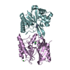
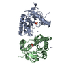
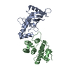
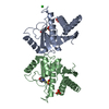
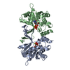
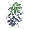
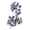
| Title | E. coli lytic transglycosylase MltA-D308A in apo-2 form | ||||||
 Components Components | Membrane-bound lytic murein transglycosylase A | ||||||
 Keywords Keywords |  HYDROLASE / double-psi beta-barrel / lytic transglycosylase / active site mutant HYDROLASE / double-psi beta-barrel / lytic transglycosylase / active site mutant | ||||||
| Function / homology |  Function and homology information Function and homology information: / lytic transglycosylase activity / peptidoglycan turnover / hydrolase activity, hydrolyzing O-glycosyl compounds / peptidoglycan catabolic process / cell wall organization / cell outer membrane / outer membrane-bounded periplasmic space Similarity search - Function | ||||||
| Biological species |  Escherichia coli (E. coli) Escherichia coli (E. coli) | ||||||
| Method | X-RAY DIFFRACTION / SYNCHROTRON / MOLECULAR REPLACEMENT / Resolution: 2.25 Å | ||||||
 Authors Authors | van Straaten, K.E. / Dijkstra, B.W. / Thunnissen, A.M.W.H. | ||||||
 Citation Citation | Journal: J.Biol.Chem. / Year: 2007 Title: Structure of Escherichia coli Lytic transglycosylase MltA with bound chitohexaose: implications for peptidoglycan binding and cleavage Authors: van Straaten, K.E. / Barends, T.R. / Dijkstra, B.W. / Thunnissen, A.M.W.H. #1: Journal: Acta Crystallogr.,Sect.D / Year: 2005Title: Escherichia coli MltA: MAD phasing and refinement of a tetartohedrally twinned protein crystal structure Authors: Barends, T.R.M. / de Jong, R.M. / van Straaten, K.E. / Thunnissen, A.M.W.H. / Dijkstra, B.W. #2: Journal: J.Mol.Biol. / Year: 2005Title: Crystal structure of MltA from Escherichia coli reveals a unique lytic transglycosylase fold Authors: van Straaten, K.E. / Dijkstra, B.W. / Vollmer, W. / Thunnissen, A.M.W.H. #3: Journal: Acta Crystallogr.,Sect.D / Year: 2004Title: Purification, crystallization and preliminary X-ray analysis of the lytic transglycosylase MltA from Escherichia coli Authors: van Straaten, K.E. / Dijkstra, B.W. / Thunnissen, A.M.W.H. | ||||||
| History |
|
- Structure visualization
| Structure viewer | Molecule: MolmilJmol/JSmol |
|---|
- Downloads & links
-Download
| PDBx/mmCIF format | 2pic.cif.gz | 82.8 KB | Display | PDBx/mmCIF format |
|---|---|---|---|---|
| PDB format | pdb2pic.ent.gz | 61.9 KB | Display | PDB format |
| PDBx/mmJSON format | 2pic.json.gz | Tree view | PDBx/mmJSON format | |
| Others |  Other downloads Other downloads |
-Validation report
| Arichive directory | https://data.pdbj.org/pub/pdb/validation_reports/pi/2picftp://data.pdbj.org/pub/pdb/validation_reports/pi/2pic | HTTPS FTP |
|---|
-Related structure data
| Related structure data | 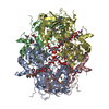 2pi8C 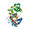 2pjjC 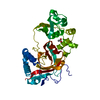 2ae0S S: Starting model for refinement C: citing same article ( |
|---|---|
| Similar structure data |

-Links
 PDBj
PDBj


- Assembly
| Deposited unit | 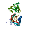
| ||||||||
|---|---|---|---|---|---|---|---|---|---|
| 1 |
| ||||||||
| Unit cell |
| ||||||||
| Components on special symmetry positions |
|
-Components
| #1: Protein | Mass: 38206.680 Da / Num. of mol.: 1 / Mutation: D308A Source method: isolated from a genetically manipulated source Source: (gene. exp.) Escherichia coli (E. coli) / Strain: K12 / Gene: mltA, mlt / Plasmid: pMSS / Species (production host): Escherichia coli / Production host: Escherichia coli BL21 (bacteria) / Strain (production host): BL21References: UniProt: P0A935, Hydrolases; Glycosylases; Glycosidases, i.e. enzymes that hydrolyse O- and S-glycosyl compounds |
|---|---|
| #2: Water | ChemComp-HOH / Water Mass: 18.015 Da / Num. of mol.: 138 / Source method: isolated from a natural source / Formula: H2O Mass: 18.015 Da / Num. of mol.: 138 / Source method: isolated from a natural source / Formula: H2O |
-Experimental details
-Experiment
| Experiment | Method: X-RAY DIFFRACTION / Number of used crystals: 1 |
|---|
- Sample preparation
| Crystal | Density Matthews: 3.22 Å3/Da / Density % sol: 61.8 % |
|---|---|
| Crystal grow | Temperature: 298 K / Method: vapor diffusion, hanging drop / pH: 4.2 Details: 0.25-0.35M NaCl, 10mM magnesium chloride, 100mM sodium acetate buffer, pH 4.2, vapor diffusion, hanging drop, temperature 298K |
-Data collection
| Diffraction | Mean temperature: 100 K | ||||||||||||||||||||||||||||||||||||||||||||||||||||||||||||||||||
|---|---|---|---|---|---|---|---|---|---|---|---|---|---|---|---|---|---|---|---|---|---|---|---|---|---|---|---|---|---|---|---|---|---|---|---|---|---|---|---|---|---|---|---|---|---|---|---|---|---|---|---|---|---|---|---|---|---|---|---|---|---|---|---|---|---|---|---|
| Diffraction source | Source: SYNCHROTRON / Site: ESRF  / Beamline: ID14-1 / Wavelength: 0.934 Å / Beamline: ID14-1 / Wavelength: 0.934 Å | ||||||||||||||||||||||||||||||||||||||||||||||||||||||||||||||||||
| Detector | Type: ADSC QUANTUM 4 / Detector: CCD | ||||||||||||||||||||||||||||||||||||||||||||||||||||||||||||||||||
| Radiation | Protocol: SINGLE WAVELENGTH / Monochromatic (M) / Laue (L): M / Scattering type: x-ray | ||||||||||||||||||||||||||||||||||||||||||||||||||||||||||||||||||
| Radiation wavelength | Wavelength: 0.934 Å / Relative weight: 1 | ||||||||||||||||||||||||||||||||||||||||||||||||||||||||||||||||||
| Reflection | Resolution: 2.25→30 Å / Num. all: 22884 / Num. obs: 22884 / % possible obs: 93.9 % / Observed criterion σ(F): 1 / Observed criterion σ(I): 1 / Redundancy: 3.6 % / Rmerge(I) obs: 0.063 / Χ2: 1.046 / Net I/σ(I): 21.5 | ||||||||||||||||||||||||||||||||||||||||||||||||||||||||||||||||||
| Reflection shell |
|
- Processing
| Software |
| ||||||||||||||||||||||||||||||||||||||||||||||||||||||||||||||||||||||
|---|---|---|---|---|---|---|---|---|---|---|---|---|---|---|---|---|---|---|---|---|---|---|---|---|---|---|---|---|---|---|---|---|---|---|---|---|---|---|---|---|---|---|---|---|---|---|---|---|---|---|---|---|---|---|---|---|---|---|---|---|---|---|---|---|---|---|---|---|---|---|---|
| Refinement | Method to determine structure: MOLECULAR REPLACEMENT Starting model: PDB ENTRY 2AE0 Resolution: 2.25→30 Å / Cor.coef. Fo:Fc: 0.943 / Cor.coef. Fo:Fc free: 0.906 / SU B: 6.265 / SU ML: 0.151 / Cross valid method: THROUGHOUT / σ(F): 0 / σ(I): 0 / ESU R: 0.245 / ESU R Free: 0.22 / Stereochemistry target values: MAXIMUM LIKELIHOOD
| ||||||||||||||||||||||||||||||||||||||||||||||||||||||||||||||||||||||
| Solvent computation | Ion probe radii: 0.8 Å / Shrinkage radii: 0.8 Å / VDW probe radii: 1.4 Å / Solvent model: BABINET MODEL WITH MASK | ||||||||||||||||||||||||||||||||||||||||||||||||||||||||||||||||||||||
| Displacement parameters | Biso mean: 49.472 Å2
| ||||||||||||||||||||||||||||||||||||||||||||||||||||||||||||||||||||||
| Refinement step | Cycle: LAST / Resolution: 2.25→30 Å
| ||||||||||||||||||||||||||||||||||||||||||||||||||||||||||||||||||||||
| Refine LS restraints |
| ||||||||||||||||||||||||||||||||||||||||||||||||||||||||||||||||||||||
| LS refinement shell | Resolution: 2.25→2.273 Å / Total num. of bins used: 50
|
