 Movie
Movie Controller

[English] 日本語
 Yorodumi
- PDB-2php: Crystal structure of the C-terminal domain of protein MJ0236 (Y23... -
+ Open data

- Basic information
| Entry | Database: PDB / ID: 2php | ||||||
|---|---|---|---|---|---|---|---|
| Title | Crystal structure of the C-terminal domain of protein MJ0236 (Y236_METJA) | ||||||
 Components Components | Uncharacterized protein MJ0236 | ||||||
 Keywords Keywords |  STRUCTURAL GENOMICS / UNKNOWN FUNCTION / Chlorine ion / PSI-2 / 10417a / NYSGXRC / Protein Structure Initiative / New York SGX Research Center for Structural Genomics STRUCTURAL GENOMICS / UNKNOWN FUNCTION / Chlorine ion / PSI-2 / 10417a / NYSGXRC / Protein Structure Initiative / New York SGX Research Center for Structural Genomics | ||||||
| Function / homology |  Function and homology informationhydroxymethylpyrimidine kinase / phosphooxymethylpyrimidine kinase / hydroxymethylpyrimidine kinase activity / thiamine phosphate synthase / phosphomethylpyrimidine kinase activity / thiamine-phosphate diphosphorylase activity / thiamine diphosphate biosynthetic process / thiamine biosynthetic process / ATP binding / cytosol / cytoplasm Function and homology informationhydroxymethylpyrimidine kinase / phosphooxymethylpyrimidine kinase / hydroxymethylpyrimidine kinase activity / thiamine phosphate synthase / phosphomethylpyrimidine kinase activity / thiamine-phosphate diphosphorylase activity / thiamine diphosphate biosynthetic process / thiamine biosynthetic process / ATP binding / cytosol / cytoplasmSimilarity search - Function | ||||||
| Biological species |   Methanocaldococcus jannaschii DSM 2661 (archaea) Methanocaldococcus jannaschii DSM 2661 (archaea) | ||||||
| Method | X-RAY DIFFRACTION / SYNCHROTRON / SAD / Resolution: 2.03 Å | ||||||
 Authors Authors | Eswaramoorthy, S. / Burley, S.K. / Swaminathan, S. / New York SGX Research Center for Structural Genomics (NYSGXRC) | ||||||
 Citation Citation | Journal: To be Published Title: Crystal structure of the C-terminal domain of protein MJ0236 (Y236_METJA) Authors: Eswaramoorthy, S. / Burley, S.K. / Swaminathan, S. | ||||||
| History |
|
- Structure visualization
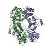
| Structure viewer | Molecule: MolmilJmol/JSmol |
|---|
- Downloads & links
-Download
| PDBx/mmCIF format | 2php.cif.gz | 154.2 KB | Display | PDBx/mmCIF format |
|---|---|---|---|---|
| PDB format | pdb2php.ent.gz | 129.5 KB | Display | PDB format |
| PDBx/mmJSON format | 2php.json.gz | Tree view | PDBx/mmJSON format | |
| Others |  Other downloads Other downloads |
-Validation report
| Arichive directory | https://data.pdbj.org/pub/pdb/validation_reports/ph/2phpftp://data.pdbj.org/pub/pdb/validation_reports/ph/2php | HTTPS FTP |
|---|
-Related structure data
| Related structure data | |
|---|---|
| Similar structure data | |
| Other databases |


-Links
 PDBj
PDBj- Assembly
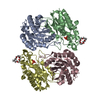
| Deposited unit | 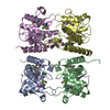
| ||||||||
|---|---|---|---|---|---|---|---|---|---|
| 1 |
| ||||||||
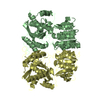
| Unit cell |
|
-Components
| #1: Protein | Mass: 21859.711 Da / Num. of mol.: 4 / Fragment: Residues 240-420 Source method: isolated from a genetically manipulated source Source: (gene. exp.) Methanocaldococcus jannaschii DSM 2661 (archaea)Species: Methanocaldococcus jannaschii / Strain: DSM 2661, JAL-1, JCM 10045, NBRC 100440 / Gene: MJ0236 / Plasmid: pSGX3(BC) / Species (production host): Escherichia coli / Production host:  Escherichia coli BL21(DE3) (bacteria) / Strain (production host): BL21(DE3) / References: UniProt: Q57688 Escherichia coli BL21(DE3) (bacteria) / Strain (production host): BL21(DE3) / References: UniProt: Q57688#2: Chemical | ChemComp-CL / Chloride  Mass: 35.453 Da / Num. of mol.: 9 / Source method: obtained synthetically / Formula: Cl Mass: 35.453 Da / Num. of mol.: 9 / Source method: obtained synthetically / Formula: Cl#3: Water | ChemComp-HOH / | Water Mass: 18.015 Da / Num. of mol.: 330 / Source method: isolated from a natural source / Formula: H2O Mass: 18.015 Da / Num. of mol.: 330 / Source method: isolated from a natural source / Formula: H2O |
|---|
-Experimental details
-Experiment
| Experiment | Method: X-RAY DIFFRACTION / Number of used crystals: 1 |
|---|
- Sample preparation
| Crystal | Density Matthews: 2.99 Å3/Da / Density % sol: 58.9 % |
|---|---|
| Crystal grow | Temperature: 295 K / Method: vapor diffusion, sitting drop / pH: 7 Details: 10% PEG6000, 2.0M Sodium chloride, pH 7.0, VAPOR DIFFUSION, SITTING DROP, temperature 295K |
-Data collection
| Diffraction | Mean temperature: 100 K |
|---|---|
| Diffraction source | Source: SYNCHROTRON / Site: NSLS  / Beamline: X12C / Wavelength: 0.9795 Å / Beamline: X12C / Wavelength: 0.9795 Å |
| Detector | Type: ADSC QUANTUM 210 / Detector: CCD / Date: Mar 26, 2007 / Details: mirrors |
| Radiation | Monochromator: Si (111) channel / Protocol: SINGLE WAVELENGTH / Monochromatic (M) / Laue (L): M / Scattering type: x-ray |
| Radiation wavelength | Wavelength: 0.9795 Å / Relative weight: 1 |
| Reflection | Resolution: 2.03→50 Å / Num. all: 63779 / Num. obs: 63779 / % possible obs: 97.2 % / Observed criterion σ(F): 0 / Observed criterion σ(I): 0 / Redundancy: 6.9 % / Rmerge(I) obs: 0.084 / Net I/σ(I): 12.8 |
| Reflection shell | Resolution: 2.03→2.1 Å / Redundancy: 5.3 % / Rmerge(I) obs: 0.413 / Num. unique all: 5451 / % possible all: 83.3 |
- Processing
| Software |
| |||||||||||||||||||||||||
|---|---|---|---|---|---|---|---|---|---|---|---|---|---|---|---|---|---|---|---|---|---|---|---|---|---|---|
| Refinement | Method to determine structure: SAD / Resolution: 2.03→50 Å / Isotropic thermal model: Isotropic / Cross valid method: THROUGHOUT / Stereochemistry target values: Engh & Huber Details: Residues listed as missing in Remark 465 are due to lack of electron density. Residues with missing atoms listed in Remark 470 are due to lack of electron density for side chains and modeled as alanines.
| |||||||||||||||||||||||||
| Displacement parameters | Biso mean: 38.9 Å2
| |||||||||||||||||||||||||
| Refine analyze |
| |||||||||||||||||||||||||
| Refinement step | Cycle: LAST / Resolution: 2.03→50 Å
| |||||||||||||||||||||||||
| Refine LS restraints |
| |||||||||||||||||||||||||
| LS refinement shell | Resolution: 2.03→2.09 Å / Rfactor Rfree error: 0.025 / Total num. of bins used: 10
|
