 Movie
Movie Controller

[English] 日本語
 Yorodumi
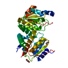
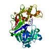
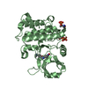
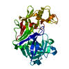
- PDB-2ph4: Crystal structure of a novel Arg49 phospholipase A2 homologue fro... -
+ Open data

- Basic information
| Entry | Database: PDB / ID: 2ph4 | ||||||
|---|---|---|---|---|---|---|---|
| Title | Crystal structure of a novel Arg49 phospholipase A2 homologue from Zhaoermia mangshanensis venom | ||||||
 Components Components | Zhaoermiatoxin | ||||||
 Keywords Keywords |  TOXIN / snake venom / arg49 / phospholipase A2 / myotoxin TOXIN / snake venom / arg49 / phospholipase A2 / myotoxin | ||||||
| Function / homology |  Function and homology informationphospholipase A2 activity / arachidonic acid secretion / phospholipid metabolic process / lipid catabolic process / toxin activity / calcium ion binding / extracellular region Function and homology informationphospholipase A2 activity / arachidonic acid secretion / phospholipid metabolic process / lipid catabolic process / toxin activity / calcium ion binding / extracellular regionSimilarity search - Function | ||||||
| Biological species |  Zhaoermia mangshanensis (snake) Zhaoermia mangshanensis (snake) | ||||||
| Method | X-RAY DIFFRACTION / SYNCHROTRON / MOLECULAR REPLACEMENT / Resolution: 2.05 Å | ||||||
 Authors Authors | Murakami, M.T. / Kuch, U. / Mebs, D. / Arni, R.K. | ||||||
 Citation Citation | Journal: Toxicon / Year: 2008 Title: Crystal structure of a novel myotoxic Arg49 phospholipase A(2) homolog (zhaoermiatoxin) from Zhaoermia mangshanensis snake venom: Insights into Arg49 coordination and the role of Lys122 in the ...Title: Crystal structure of a novel myotoxic Arg49 phospholipase A(2) homolog (zhaoermiatoxin) from Zhaoermia mangshanensis snake venom: Insights into Arg49 coordination and the role of Lys122 in the polarization of the C-terminus. Authors: Murakami, M.T. / Kuch, U. / Betzel, C. / Mebs, D. / Arni, R.K. | ||||||
| History |
| ||||||
| Remark 999 | sequence THE SEQUENCE IS NUMBERED BASED ON THE BOVINE PANCREATIC PHOSPHOLIPASE A2. SO, NOT ...sequence THE SEQUENCE IS NUMBERED BASED ON THE BOVINE PANCREATIC PHOSPHOLIPASE A2. SO, NOT SEQUENTIAL NUMBERS INDICATE DELETIONS AND INSERTIONS REGIONS IN COMPARISON WITH BOVINE PANCREATIC PLA2. |
- Structure visualization
| Structure viewer | Molecule: MolmilJmol/JSmol |
|---|
- Downloads & links
-Download
| PDBx/mmCIF format | 2ph4.cif.gz | 67.4 KB | Display | PDBx/mmCIF format |
|---|---|---|---|---|
| PDB format | pdb2ph4.ent.gz | 50.3 KB | Display | PDB format |
| PDBx/mmJSON format | 2ph4.json.gz | Tree view | PDBx/mmJSON format | |
| Others |  Other downloads Other downloads |
-Validation report
| Arichive directory | https://data.pdbj.org/pub/pdb/validation_reports/ph/2ph4ftp://data.pdbj.org/pub/pdb/validation_reports/ph/2ph4 | HTTPS FTP |
|---|
-Related structure data
| Related structure data |  1y4lS S: Starting model for refinement |
|---|---|
| Similar structure data |


-Links
 PDBj
PDBj
- Assembly
| Deposited unit | 
| ||||||||
|---|---|---|---|---|---|---|---|---|---|
| 1 |
| ||||||||
| 2 | 
| ||||||||
| Unit cell |
| ||||||||
| Details | The biological assembly is the dimer found in the asymmetric unit. |
-Components
| #1: Protein | Mass: 14010.464 Da / Num. of mol.: 2 / Source method: isolated from a natural source / Source: (natural) Zhaoermia mangshanensis (snake) / Secretion: venom / References: UniProt: P84776#2: Chemical | ChemComp-SO4 / Sulfate  Mass: 96.063 Da / Num. of mol.: 5 / Source method: obtained synthetically / Formula: SO4 Mass: 96.063 Da / Num. of mol.: 5 / Source method: obtained synthetically / Formula: SO4#3: Chemical | Diethylene glycol  Mass: 106.120 Da / Num. of mol.: 2 / Source method: obtained synthetically / Formula: C4H10O3 Mass: 106.120 Da / Num. of mol.: 2 / Source method: obtained synthetically / Formula: C4H10O3#4: Water | ChemComp-HOH / | Water Mass: 18.015 Da / Num. of mol.: 235 / Source method: isolated from a natural source / Formula: H2O Mass: 18.015 Da / Num. of mol.: 235 / Source method: isolated from a natural source / Formula: H2O |
|---|
-Experimental details
-Experiment
| Experiment | Method: X-RAY DIFFRACTION / Number of used crystals: 1 |
|---|
- Sample preparation
| Crystal | Density Matthews: 2.57 Å3/Da / Density % sol: 52.19 % |
|---|---|
| Crystal grow | Temperature: 291 K / Method: vapor diffusion, hanging drop / pH: 6.5 Details: polyethylene glycol 8,000; ammonium sulfate, pH 6.5, VAPOR DIFFUSION, HANGING DROP, temperature 291K |
-Data collection
| Diffraction | Mean temperature: 100 K |
|---|---|
| Diffraction source | Source: SYNCHROTRON / Site: LNLS  / Beamline: D03B-MX1 / Wavelength: 1.427 Å / Beamline: D03B-MX1 / Wavelength: 1.427 Å |
| Detector | Type: MAR CCD 165 mm / Detector: CCD / Date: Feb 10, 2007 |
| Radiation | Protocol: SINGLE WAVELENGTH / Monochromatic (M) / Laue (L): M / Scattering type: x-ray |
| Radiation wavelength | Wavelength: 1.427 Å / Relative weight: 1 |
| Reflection | Resolution: 2.05→23.87 Å / Num. obs: 17731 / % possible obs: 99.5 % / Observed criterion σ(I): 2 / Redundancy: 12.5 % / Rmerge(I) obs: 0.071 / Net I/σ(I): 33.4 |
| Reflection shell | Resolution: 2.05→2.12 Å / Redundancy: 9.5 % / Rmerge(I) obs: 0.33 / Mean I/σ(I) obs: 2.8 / % possible all: 99.4 |
- Processing
| Software |
| ||||||||||||||||||||||||||||||||||||||||||||||||||||||||||||||||||||||||||||||||||||||||||
|---|---|---|---|---|---|---|---|---|---|---|---|---|---|---|---|---|---|---|---|---|---|---|---|---|---|---|---|---|---|---|---|---|---|---|---|---|---|---|---|---|---|---|---|---|---|---|---|---|---|---|---|---|---|---|---|---|---|---|---|---|---|---|---|---|---|---|---|---|---|---|---|---|---|---|---|---|---|---|---|---|---|---|---|---|---|---|---|---|---|---|---|
| Refinement | Method to determine structure: MOLECULAR REPLACEMENT Starting model: PDB ENTRY 1Y4L Resolution: 2.05→23.87 Å / Cor.coef. Fo:Fc: 0.942 / Cor.coef. Fo:Fc free: 0.883 / SU B: 5.84 / SU ML: 0.16 / Isotropic thermal model: isotropic / Cross valid method: THROUGHOUT / ESU R: 0.25 / ESU R Free: 0.229 / Stereochemistry target values: MAXIMUM LIKELIHOOD / Details: HYDROGENS HAVE BEEN ADDED IN THE RIDING POSITIONS
| ||||||||||||||||||||||||||||||||||||||||||||||||||||||||||||||||||||||||||||||||||||||||||
| Solvent computation | Ion probe radii: 0.8 Å / Shrinkage radii: 0.8 Å / VDW probe radii: 1.4 Å / Solvent model: MASK | ||||||||||||||||||||||||||||||||||||||||||||||||||||||||||||||||||||||||||||||||||||||||||
| Displacement parameters | Biso mean: 29.077 Å2
| ||||||||||||||||||||||||||||||||||||||||||||||||||||||||||||||||||||||||||||||||||||||||||
| Refinement step | Cycle: LAST / Resolution: 2.05→23.87 Å
| ||||||||||||||||||||||||||||||||||||||||||||||||||||||||||||||||||||||||||||||||||||||||||
| Refine LS restraints |
| ||||||||||||||||||||||||||||||||||||||||||||||||||||||||||||||||||||||||||||||||||||||||||
| LS refinement shell | Resolution: 2.05→2.103 Å / Total num. of bins used: 20
|
