 Movie
Movie Controller

[English] 日本語
 Yorodumi
- PDB-2pfs: Crystal structure of universal stress protein from Nitrosomonas e... -
+ Open data

- Basic information
| Entry | Database: PDB / ID: 2pfs | ||||||
|---|---|---|---|---|---|---|---|
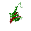
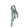
| Title | Crystal structure of universal stress protein from Nitrosomonas europaea | ||||||
 Components Components | Universal stress protein | ||||||
 Keywords Keywords | STRUCTURAL GENOMICS / UNKNOWN FUNCTION / stress protein / PSI-2 / Protein Structure Initiative / Midwest Center for Structural Genomics / MCSG | ||||||
| Function / homology |  Function and homology information Function and homology information | ||||||
| Biological species |  Nitrosomonas europaea ATCC 19718 (bacteria) Nitrosomonas europaea ATCC 19718 (bacteria) | ||||||
| Method | X-RAY DIFFRACTION / SYNCHROTRON / SAD / Resolution: 2.25 Å | ||||||
 Authors Authors | Chruszcz, M. / Evdokimova, E. / Cymborowski, M. / Kagan, O. / Savchenko, A. / Edwards, A. / Joachimiak, A. / Minor, W. / Midwest Center for Structural Genomics (MCSG) | ||||||
 Citation Citation | Journal: Evol Appl / Year: 2013 Title: Structural and functional insight into the universal stress protein family. Authors: Tkaczuk, K.L. / Shumilin, I.A. / Chruszcz, M. / Evdokimova, E. / Savchenko, A. / Minor, W. | ||||||
| History |
|
- Structure visualization
| Structure viewer | Molecule: MolmilJmol/JSmol |
|---|
- Downloads & links
-Download
| PDBx/mmCIF format | 2pfs.cif.gz | 35.7 KB | Display | PDBx/mmCIF format |
|---|---|---|---|---|
| PDB format | pdb2pfs.ent.gz | 26.6 KB | Display | PDB format |
| PDBx/mmJSON format | 2pfs.json.gz | Tree view | PDBx/mmJSON format | |
| Others |  Other downloads Other downloads |
-Validation report
| Arichive directory | https://data.pdbj.org/pub/pdb/validation_reports/pf/2pfsftp://data.pdbj.org/pub/pdb/validation_reports/pf/2pfs | HTTPS FTP |
|---|
-Related structure data
| Related structure data |  3qtbC  3tnjC 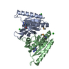 6hcdC C: citing same article ( |
|---|---|
| Similar structure data | |
| Other databases |


-Links
 PDBj
PDBj- Assembly
| Deposited unit | 
| ||||||||
|---|---|---|---|---|---|---|---|---|---|
| 1 | 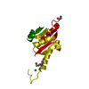
| ||||||||
| Unit cell |
|
-Components
| #1: Protein | / Usp Mass: 16756.553 Da / Num. of mol.: 1 Source method: isolated from a genetically manipulated source Source: (gene. exp.) Nitrosomonas europaea ATCC 19718 (bacteria)Species: Nitrosomonas europaea / Strain: IFO 14298 / Gene: NE1028 / Plasmid: p15TV-L / Production host: Escherichia coli (E. coli) / Strain (production host): BL21-Gold(DE3) / References: UniProt: Q82VN8 | ||
|---|---|---|---|
| #2: Chemical | Chloride  Mass: 35.453 Da / Num. of mol.: 2 / Source method: obtained synthetically / Formula: Cl Mass: 35.453 Da / Num. of mol.: 2 / Source method: obtained synthetically / Formula: Cl#3: Water | ChemComp-HOH / | Water Mass: 18.015 Da / Num. of mol.: 40 / Source method: isolated from a natural source / Formula: H2O Mass: 18.015 Da / Num. of mol.: 40 / Source method: isolated from a natural source / Formula: H2O |
-Experimental details
-Experiment
| Experiment | Method: X-RAY DIFFRACTION / Number of used crystals: 1 |
|---|
- Sample preparation
| Crystal | Density Matthews: 2.14 Å3/Da / Density % sol: 42.47 % |
|---|---|
| Crystal grow | Temperature: 293 K / Method: vapor diffusion / pH: 7.5 Details: 0.2M Sodium chloride, 0.1M HEPES pH 7.5, 25% PEG3350, 2% Isopropanol, VAPOR DIFFUSION, temperature 293K |
-Data collection
| Diffraction | Mean temperature: 100 K |
|---|---|
| Diffraction source | Source: SYNCHROTRON / Site: APS  / Beamline: 19-ID / Wavelength: 0.97918 Å / Beamline: 19-ID / Wavelength: 0.97918 Å |
| Detector | Type: ADSC QUANTUM 315 / Detector: CCD / Date: Feb 8, 2007 / Details: Mirrors |
| Radiation | Monochromator: Si 111 CHANNEL / Protocol: SINGLE WAVELENGTH / Monochromatic (M) / Laue (L): M / Scattering type: x-ray |
| Radiation wavelength | Wavelength: 0.97918 Å / Relative weight: 1 |
| Reflection | Resolution: 2.25→50 Å / Num. all: 6882 / Num. obs: 6882 / % possible obs: 97.6 % / Observed criterion σ(F): 0 / Observed criterion σ(I): -3 / Redundancy: 11.1 % / Rmerge(I) obs: 0.06 / Rsym value: 0.06 / Net I/σ(I): 58.5 |
| Reflection shell | Resolution: 2.25→2.29 Å / Redundancy: 7.7 % / Rmerge(I) obs: 0.486 / Mean I/σ(I) obs: 2.9 / Num. unique all: 277 / Rsym value: 0.486 / % possible all: 81.5 |
- Processing
| Software |
| ||||||||||||||||||||||||||||||||||||||||||||||||||||||||||||||||||||||||||||||||||||||||||||||||||||||||||||||||||||||||||||||||||||||||||||||||||||||||||||||||||||||||||
|---|---|---|---|---|---|---|---|---|---|---|---|---|---|---|---|---|---|---|---|---|---|---|---|---|---|---|---|---|---|---|---|---|---|---|---|---|---|---|---|---|---|---|---|---|---|---|---|---|---|---|---|---|---|---|---|---|---|---|---|---|---|---|---|---|---|---|---|---|---|---|---|---|---|---|---|---|---|---|---|---|---|---|---|---|---|---|---|---|---|---|---|---|---|---|---|---|---|---|---|---|---|---|---|---|---|---|---|---|---|---|---|---|---|---|---|---|---|---|---|---|---|---|---|---|---|---|---|---|---|---|---|---|---|---|---|---|---|---|---|---|---|---|---|---|---|---|---|---|---|---|---|---|---|---|---|---|---|---|---|---|---|---|---|---|---|---|---|---|---|---|---|
| Refinement | Method to determine structure: SAD / Resolution: 2.25→50 Å / Cor.coef. Fo:Fc: 0.951 / Cor.coef. Fo:Fc free: 0.927 / SU B: 11.231 / SU ML: 0.141 / TLS residual ADP flag: LIKELY RESIDUAL / Cross valid method: THROUGHOUT / σ(F): 0 / σ(I): 0 / ESU R: 0.277 / ESU R Free: 0.228 / Stereochemistry target values: MAXIMUM LIKELIHOOD / Details: HYDROGENS HAVE BEEN ADDED IN THE RIDING POSITIONS
| ||||||||||||||||||||||||||||||||||||||||||||||||||||||||||||||||||||||||||||||||||||||||||||||||||||||||||||||||||||||||||||||||||||||||||||||||||||||||||||||||||||||||||
| Solvent computation | Ion probe radii: 0.8 Å / Shrinkage radii: 0.8 Å / VDW probe radii: 1.2 Å / Solvent model: MASK | ||||||||||||||||||||||||||||||||||||||||||||||||||||||||||||||||||||||||||||||||||||||||||||||||||||||||||||||||||||||||||||||||||||||||||||||||||||||||||||||||||||||||||
| Displacement parameters | Biso mean: 52.42 Å2
| ||||||||||||||||||||||||||||||||||||||||||||||||||||||||||||||||||||||||||||||||||||||||||||||||||||||||||||||||||||||||||||||||||||||||||||||||||||||||||||||||||||||||||
| Refinement step | Cycle: LAST / Resolution: 2.25→50 Å
| ||||||||||||||||||||||||||||||||||||||||||||||||||||||||||||||||||||||||||||||||||||||||||||||||||||||||||||||||||||||||||||||||||||||||||||||||||||||||||||||||||||||||||
| Refine LS restraints |
| ||||||||||||||||||||||||||||||||||||||||||||||||||||||||||||||||||||||||||||||||||||||||||||||||||||||||||||||||||||||||||||||||||||||||||||||||||||||||||||||||||||||||||
| LS refinement shell | Resolution: 2.25→2.309 Å / Total num. of bins used: 20
| ||||||||||||||||||||||||||||||||||||||||||||||||||||||||||||||||||||||||||||||||||||||||||||||||||||||||||||||||||||||||||||||||||||||||||||||||||||||||||||||||||||||||||
| Refinement TLS params. | Method: refined / Refine-ID: X-RAY DIFFRACTION
| ||||||||||||||||||||||||||||||||||||||||||||||||||||||||||||||||||||||||||||||||||||||||||||||||||||||||||||||||||||||||||||||||||||||||||||||||||||||||||||||||||||||||||
| Refinement TLS group |
|
