 Movie
Movie Controller

[English] 日本語
 Yorodumi
- PDB-2pfi: Crystal structure of the cytoplasmic domain of the human chloride... -
+ Open data

- Basic information
| Entry | Database: PDB / ID: 2pfi | ||||||
|---|---|---|---|---|---|---|---|
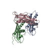
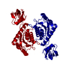
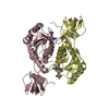
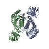
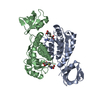
| Title | Crystal structure of the cytoplasmic domain of the human chloride channel ClC-Ka | ||||||
 Components Components | Chloride channel protein ClC-Ka | ||||||
 Keywords Keywords | TRANSPORT PROTEIN / Cystathionine beta synthetase (CBS) domains containing protein | ||||||
| Function / homology |  Function and homology informationexcretion / voltage-gated chloride channel activity / regulation of monoatomic ion transmembrane transport / chloride transport / plasma membrane => GO:0005886 / chloride channel complex / monoatomic ion transmembrane transport / Stimuli-sensing channels / identical protein binding / metal ion binding / plasma membrane Function and homology informationexcretion / voltage-gated chloride channel activity / regulation of monoatomic ion transmembrane transport / chloride transport / plasma membrane => GO:0005886 / chloride channel complex / monoatomic ion transmembrane transport / Stimuli-sensing channels / identical protein binding / metal ion binding / plasma membraneSimilarity search - Function | ||||||
| Biological species |  Homo sapiens (human) Homo sapiens (human) | ||||||
| Method | X-RAY DIFFRACTION / SYNCHROTRON / MIR / Resolution: 1.6 Å | ||||||
 Authors Authors | Markovic, S. / Dutzler, R. | ||||||
 Citation Citation | Journal: Structure / Year: 2007 Title: The Structure of the Cytoplasmic Domain of the Chloride Channel ClC-Ka Reveals a Conserved Interaction Interface. Authors: Markovic, S. / Dutzler, R. | ||||||
| History |
| ||||||
| Remark 999 | SEQUENCE THE SEQUENCE WAS CLONED WITH C-TERMINAL RECOGNITION SITE FOR PRESCISSION PROTEASE FOLLOWED ...SEQUENCE THE SEQUENCE WAS CLONED WITH C-TERMINAL RECOGNITION SITE FOR PRESCISSION PROTEASE FOLLOWED BY A HEXA-HISTIDINE TAG THAT WAS CLEAVED OFF DURING PURIFICATION |
- Structure visualization
| Structure viewer | Molecule: MolmilJmol/JSmol |
|---|
- Downloads & links
-Download
| PDBx/mmCIF format | 2pfi.cif.gz | 75.8 KB | Display | PDBx/mmCIF format |
|---|---|---|---|---|
| PDB format | pdb2pfi.ent.gz | 56.3 KB | Display | PDB format |
| PDBx/mmJSON format | 2pfi.json.gz | Tree view | PDBx/mmJSON format | |
| Others |  Other downloads Other downloads |
-Validation report
| Arichive directory | https://data.pdbj.org/pub/pdb/validation_reports/pf/2pfiftp://data.pdbj.org/pub/pdb/validation_reports/pf/2pfi | HTTPS FTP |
|---|
-Related structure data
| Similar structure data |
|---|
-Links
 PDBj
PDBj
- Assembly
| Deposited unit | 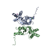
| ||||||||
|---|---|---|---|---|---|---|---|---|---|
| 1 |
| ||||||||
| Unit cell |
|
-Components
| #1: Protein | / Chloride channel Ka / ClC-K1 Mass: 18149.816 Da / Num. of mol.: 2 / Fragment: human ClC-Ka C-terminal domain Source method: isolated from a genetically manipulated source Source: (gene. exp.) Homo sapiens (human) / Gene: CLCNKA / Plasmid: pet 28b+ / Species (production host): Escherichia coli / Production host:  Escherichia coli BL21(DE3) (bacteria) / Strain (production host): Bl21/DE3 / References: UniProt: P51800 Escherichia coli BL21(DE3) (bacteria) / Strain (production host): Bl21/DE3 / References: UniProt: P51800#2: Chemical | ChemComp-IOD / | Iodide  Mass: 126.904 Da / Num. of mol.: 1 / Source method: obtained synthetically / Formula: I Mass: 126.904 Da / Num. of mol.: 1 / Source method: obtained synthetically / Formula: I#3: Chemical | Chloride  Mass: 35.453 Da / Num. of mol.: 3 / Source method: obtained synthetically / Formula: Cl Mass: 35.453 Da / Num. of mol.: 3 / Source method: obtained synthetically / Formula: Cl#4: Water | ChemComp-HOH / | Water Mass: 18.015 Da / Num. of mol.: 286 / Source method: isolated from a natural source / Formula: H2O Mass: 18.015 Da / Num. of mol.: 286 / Source method: isolated from a natural source / Formula: H2O |
|---|
-Experimental details
-Experiment
| Experiment | Method: X-RAY DIFFRACTION / Number of used crystals: 1 |
|---|
- Sample preparation
| Crystal | Density Matthews: 2.47 Å3/Da / Density % sol: 50.19 % |
|---|---|
| Crystal grow | Temperature: 277 K / Method: vapor diffusion, sitting drop / pH: 8.5 Details: 20% PEG 4000, 100mM KI, 60mM Tris-HCl, 150mM NaCl, pH 8.5, VAPOR DIFFUSION, SITTING DROP, temperature 277.0K |
-Data collection
| Diffraction | Mean temperature: 100 K |
|---|---|
| Diffraction source | Source: SYNCHROTRON / Site: SLS  / Beamline: X06SA / Wavelength: 1 Å / Beamline: X06SA / Wavelength: 1 Å |
| Detector | Type: MARMOSAIC 225 mm CCD / Detector: CCD / Date: Mar 27, 2006 |
| Radiation | Monochromator: Si 111 / Protocol: SINGLE WAVELENGTH / Monochromatic (M) / Laue (L): M / Scattering type: x-ray |
| Radiation wavelength | Wavelength: 1 Å / Relative weight: 1 |
| Reflection | Resolution: 1.6→50 Å / Num. all: 45049 / Num. obs: 43653 / % possible obs: 96.9 % / Observed criterion σ(F): 2 / Observed criterion σ(I): 2 / Redundancy: 3.2 % / Biso Wilson estimate: 26.4 Å2 / Rmerge(I) obs: 0.055 / Net I/σ(I): 26.9 |
| Reflection shell | Resolution: 1.6→1.64 Å / Redundancy: 1.6 % / Rmerge(I) obs: 0.278 / Mean I/σ(I) obs: 2.8 / Num. unique all: 2317 / % possible all: 79 |
- Processing
| Software |
| ||||||||||||||||||||||||||||||||||||
|---|---|---|---|---|---|---|---|---|---|---|---|---|---|---|---|---|---|---|---|---|---|---|---|---|---|---|---|---|---|---|---|---|---|---|---|---|---|
| Refinement | Method to determine structure: MIR / Resolution: 1.6→14.89 Å / Rfactor Rfree error: 0.003 / Data cutoff high absF: 820085.84 / Data cutoff low absF: 0 / Isotropic thermal model: RESTRAINED / Cross valid method: THROUGHOUT / σ(F): 0 / Stereochemistry target values: Engh & Huber
| ||||||||||||||||||||||||||||||||||||
| Solvent computation | Solvent model: FLAT MODEL / Bsol: 81.979 Å2 / ksol: 0.406422 e/Å3 | ||||||||||||||||||||||||||||||||||||
| Displacement parameters | Biso mean: 30 Å2
| ||||||||||||||||||||||||||||||||||||
| Refine analyze |
| ||||||||||||||||||||||||||||||||||||
| Refinement step | Cycle: LAST / Resolution: 1.6→14.89 Å
| ||||||||||||||||||||||||||||||||||||
| Refine LS restraints |
| ||||||||||||||||||||||||||||||||||||
| LS refinement shell | Resolution: 1.6→1.7 Å / Rfactor Rfree error: 0.013 / Total num. of bins used: 6
| ||||||||||||||||||||||||||||||||||||
| Xplor file |
|
