 Movie
Movie Controller

[English] 日本語
 Yorodumi
- PDB-2pb1: Exo-B-(1,3)-Glucanase from Candida Albicans in complex with unhyd... -
+ Open data

- Basic information
| Entry | Database: PDB / ID: 2pb1 | ||||||
|---|---|---|---|---|---|---|---|
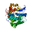
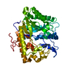
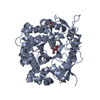
| Title | Exo-B-(1,3)-Glucanase from Candida Albicans in complex with unhydrolysed and covalently linked 2,4-dinitrophenyl-2-deoxy-2-fluoro-B-D-glucopyranoside at 1.9 A | ||||||
 Components Components | Hypothetical protein XOG1 Hypothesis Hypothesis | ||||||
 Keywords Keywords | HYDROLASE / EXO-GLUCANASE / CANDIDA ALBICANS / MECHANISM-BASED INHIBITOR | ||||||
| Function / homology |  Function and homology information Function and homology informationglucan metabolic process / single-species biofilm formation in or on host organism / glucan 1,3-beta-glucosidase / glucan exo-1,3-beta-glucosidase activity / single-species biofilm formation on inanimate substrate / fungal-type cell wall organization / glucan catabolic process / Transferases; Glycosyltransferases; Hexosyltransferases / cell-substrate adhesion / cell adhesion molecule binding ...glucan metabolic process / single-species biofilm formation in or on host organism / glucan 1,3-beta-glucosidase / glucan exo-1,3-beta-glucosidase activity / single-species biofilm formation on inanimate substrate / fungal-type cell wall organization / glucan catabolic process / Transferases; Glycosyltransferases; Hexosyltransferases / cell-substrate adhesion / cell adhesion molecule binding / extracellular vesicle / transferase activity / cell surface / extracellular regionSimilarity search - Function | ||||||
| Biological species |  Candida albicans (yeast) Candida albicans (yeast) | ||||||
| Method | X-RAY DIFFRACTION / MOLECULAR REPLACEMENT / Resolution: 1.9 Å | ||||||
 Authors Authors | Cutfield, S.M. / Cutfield, J.F. / Davies, G.J. / Sullivan, P.A. | ||||||
 Citation Citation | Journal: To be Published Title: Exo-B-(1,3)-Glucanase from Candida Albicans in complex with unhydrolysed and covalently linked 2,4-dinitrophenyl-2-deoxy-2-fluoro-B-D-glucopyranoside at 1.9 A Authors: Cutfield, S.M. / Cutfield, J.F. / Davies, G.J. / Sullivan, P.A. #1: Journal: J.Mol.Biol.Title: The structure of the exo-beta-(1,3)-glucanase from Candida albicans in native and bound forms: relationship between a pocket and groove in family 5 glycosyl hydrolases. Authors: Cutfield, S.M. / Davies, G.J. / Murshudov, G. / Anderson, B.F. / Moody, P.C. / Sullivan, P.A. / Cutfield, J.F. | ||||||
| History |
| ||||||
| Remark 999 | sequence Due to alternative codon usage by Candida albicans, residue 64 is a Ser when from natural ...sequence Due to alternative codon usage by Candida albicans, residue 64 is a Ser when from natural sources, and a Leu when expressed in Saccharomyces cerevisiae. |
- Structure visualization
| Structure viewer | Molecule: MolmilJmol/JSmol |
|---|
- Downloads & links
-Download
| PDBx/mmCIF format | 2pb1.cif.gz | 100 KB | Display | PDBx/mmCIF format |
|---|---|---|---|---|
| PDB format | pdb2pb1.ent.gz | 75.6 KB | Display | PDB format |
| PDBx/mmJSON format | 2pb1.json.gz | Tree view | PDBx/mmJSON format | |
| Others |  Other downloads Other downloads |
-Validation report
| Arichive directory | https://data.pdbj.org/pub/pdb/validation_reports/pb/2pb1ftp://data.pdbj.org/pub/pdb/validation_reports/pb/2pb1 | HTTPS FTP |
|---|
-Related structure data
| Related structure data | 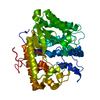 1eqpS S: Starting model for refinement |
|---|---|
| Similar structure data |


-Links
 PDBj
PDBj- Assembly
| Deposited unit | 
| ||||||||
|---|---|---|---|---|---|---|---|---|---|
| 1 |
| ||||||||
| Unit cell |
|
-Components
| #1: Protein | Hypothesis Mass: 45792.398 Da / Num. of mol.: 1 Source method: isolated from a genetically manipulated source Source: (gene. exp.) Candida albicans (yeast) / Strain: ATCC 10261 / Gene: EXG / Plasmid: pEMBLYEX4 / Production host: Saccharomyces cerevisiae (brewer's yeast) / Strain (production host): AWY-1References: UniProt: Q5AIZ3, UniProt: P29717*PLUS, glucan 1,3-beta-glucosidase |
|---|---|
| #2: Sugar | ChemComp-G2F /   Type: D-saccharide, alpha linking / Mass: 182.147 Da / Num. of mol.: 1 Type: D-saccharide, alpha linking / Mass: 182.147 Da / Num. of mol.: 1Source method: isolated from a genetically manipulated source Formula: C6H11FO5 |
| #3: Sugar | ChemComp-NFG /   Type: D-saccharide / Mass: 348.238 Da / Num. of mol.: 1 / Source method: obtained synthetically / Formula: C12H13FN2O9 Type: D-saccharide / Mass: 348.238 Da / Num. of mol.: 1 / Source method: obtained synthetically / Formula: C12H13FN2O9 |
| #4: Water | ChemComp-HOH / Water Mass: 18.015 Da / Num. of mol.: 274 / Source method: isolated from a natural source / Formula: H2O Mass: 18.015 Da / Num. of mol.: 274 / Source method: isolated from a natural source / Formula: H2O |
-Experimental details
-Experiment
| Experiment | Method: X-RAY DIFFRACTION / Number of used crystals: 1 |
|---|
- Sample preparation
| Crystal | Density Matthews: 2.09 Å3/Da / Density % sol: 41.08 % |
|---|---|
| Crystal grow | Temperature: 293 K / Method: vapor diffusion, hanging drop / pH: 7.3 Details: PEG 8000, Hepes, CaCl2, pH 7.3, temperature 293K, VAPOR DIFFUSION, HANGING DROP |
-Data collection
| Diffraction | Mean temperature: 293 K | ||||||||||||||||||||||||||||||||||||||||||||||||||||||||||||||||||
|---|---|---|---|---|---|---|---|---|---|---|---|---|---|---|---|---|---|---|---|---|---|---|---|---|---|---|---|---|---|---|---|---|---|---|---|---|---|---|---|---|---|---|---|---|---|---|---|---|---|---|---|---|---|---|---|---|---|---|---|---|---|---|---|---|---|---|---|
| Diffraction source | Source: ROTATING ANODE / Type: RIGAKU RU200 / Wavelength: 1.5418 Å | ||||||||||||||||||||||||||||||||||||||||||||||||||||||||||||||||||
| Detector | Type: RIGAKU RAXIS II / Detector: IMAGE PLATE / Date: Feb 6, 1997 | ||||||||||||||||||||||||||||||||||||||||||||||||||||||||||||||||||
| Radiation | Protocol: SINGLE WAVELENGTH / Monochromatic (M) / Laue (L): M / Scattering type: x-ray | ||||||||||||||||||||||||||||||||||||||||||||||||||||||||||||||||||
| Radiation wavelength | Wavelength: 1.5418 Å / Relative weight: 1 | ||||||||||||||||||||||||||||||||||||||||||||||||||||||||||||||||||
| Reflection | Resolution: 1.9→20 Å / Num. all: 30898 / Num. obs: 28359 / % possible obs: 91.6 % / Rmerge(I) obs: 0.047 / Χ2: 0.994 / Net I/σ(I): 14.8 | ||||||||||||||||||||||||||||||||||||||||||||||||||||||||||||||||||
| Reflection shell |
|
- Processing
| Software |
| |||||||||||||||||||||||||||||||||||||||||||||||||||||||||||||||||||||||||||||||||||||||||||||||||||||||||||||||||||||||||||||
|---|---|---|---|---|---|---|---|---|---|---|---|---|---|---|---|---|---|---|---|---|---|---|---|---|---|---|---|---|---|---|---|---|---|---|---|---|---|---|---|---|---|---|---|---|---|---|---|---|---|---|---|---|---|---|---|---|---|---|---|---|---|---|---|---|---|---|---|---|---|---|---|---|---|---|---|---|---|---|---|---|---|---|---|---|---|---|---|---|---|---|---|---|---|---|---|---|---|---|---|---|---|---|---|---|---|---|---|---|---|---|---|---|---|---|---|---|---|---|---|---|---|---|---|---|---|---|
| Refinement | Method to determine structure: MOLECULAR REPLACEMENT Starting model: PDB entry 1EQP Resolution: 1.9→19.92 Å / Cor.coef. Fo:Fc: 0.975 / Cor.coef. Fo:Fc free: 0.967 / SU B: 2.32 / SU ML: 0.07 / Cross valid method: THROUGHOUT / σ(F): 0 / ESU R: 0.139 / ESU R Free: 0.116 / Stereochemistry target values: MAXIMUM LIKELIHOOD / Details: HYDROGENS HAVE BEEN ADDED IN THE RIDING POSITIONS
| |||||||||||||||||||||||||||||||||||||||||||||||||||||||||||||||||||||||||||||||||||||||||||||||||||||||||||||||||||||||||||||
| Solvent computation | Ion probe radii: 0.8 Å / Shrinkage radii: 0.8 Å / VDW probe radii: 1.2 Å / Solvent model: MASK | |||||||||||||||||||||||||||||||||||||||||||||||||||||||||||||||||||||||||||||||||||||||||||||||||||||||||||||||||||||||||||||
| Displacement parameters | Biso mean: 20.534 Å2
| |||||||||||||||||||||||||||||||||||||||||||||||||||||||||||||||||||||||||||||||||||||||||||||||||||||||||||||||||||||||||||||
| Refinement step | Cycle: LAST / Resolution: 1.9→19.92 Å
| |||||||||||||||||||||||||||||||||||||||||||||||||||||||||||||||||||||||||||||||||||||||||||||||||||||||||||||||||||||||||||||
| Refine LS restraints |
| |||||||||||||||||||||||||||||||||||||||||||||||||||||||||||||||||||||||||||||||||||||||||||||||||||||||||||||||||||||||||||||
| LS refinement shell | Resolution: 1.9→1.949 Å / Total num. of bins used: 20
|
