 Movie
Movie Controller


+ Open data

- Basic information
| Entry | Database: PDB / ID: 2p68 | ||||||
|---|---|---|---|---|---|---|---|
| Title | Crystal Structure of aq_1716 from Aquifex Aeolicus VF5 | ||||||
 Components Components | 3-oxoacyl-[acyl-carrier-protein] reductase | ||||||
 Keywords Keywords |  OXIDOREDUCTASE / aq_1716 / 3-ketoacyl-acyl carrier protein reductase / 3-oxoacyl-[acyl-carrier-protein] reductase / Aquifex aeolicus VF5 / structural genomics / PSI / Protein Structure Initiative / Southeast Collaboratory for Structural Genomics / SECSG / NPPSFA / National Project on Protein Structural and Functional Analyses / RIKEN STRUCTURAL GENOMICS/PROTEOMICS INITIATIVE / RSGI OXIDOREDUCTASE / aq_1716 / 3-ketoacyl-acyl carrier protein reductase / 3-oxoacyl-[acyl-carrier-protein] reductase / Aquifex aeolicus VF5 / structural genomics / PSI / Protein Structure Initiative / Southeast Collaboratory for Structural Genomics / SECSG / NPPSFA / National Project on Protein Structural and Functional Analyses / RIKEN STRUCTURAL GENOMICS/PROTEOMICS INITIATIVE / RSGI | ||||||
| Function / homology |  Function and homology information Function and homology information3-oxoacyl-[acyl-carrier-protein] reductase / 3-oxoacyl-[acyl-carrier-protein] reductase (NADPH) activity / fatty acid elongation / oxidoreductase activity, acting on the CH-OH group of donors, NAD or NADP as acceptor / NAD binding / NADP bindingSimilarity search - Function | ||||||
| Biological species |   Aquifex aeolicus (bacteria) Aquifex aeolicus (bacteria) | ||||||
| Method | X-RAY DIFFRACTION / SYNCHROTRON / MOLECULAR REPLACEMENT / Resolution: 1.84 Å | ||||||
 Authors Authors | Chen, L. / Chen, L.-Q. / Ebihara, A. / Shinkai, A. / Kuramitsu, S. / Yokoyama, S. / Zhao, M. / Dillard, B. / Rose, J.P. / Wang, B.-C. ...Chen, L. / Chen, L.-Q. / Ebihara, A. / Shinkai, A. / Kuramitsu, S. / Yokoyama, S. / Zhao, M. / Dillard, B. / Rose, J.P. / Wang, B.-C. / Southeast Collaboratory for Structural Genomics (SECSG) / RIKEN Structural Genomics/Proteomics Initiative (RSGI) | ||||||
 Citation Citation | Journal: To be Published Title: Crystal Structure of aq_1716 from Aquifex aeolicus VF5 Authors: Chen, L. / Chen, L.-Q. / Ebihara, A. / Shinkai, A. / Kuramitsu, S. / Yokoyama, S. / Zhao, M. / Dillard, B. / Rose, J.P. / Wang, B.-C. | ||||||
| History |
|
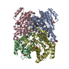
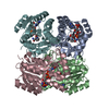
- Structure visualization
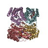
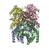
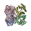
| Structure viewer | Molecule: MolmilJmol/JSmol |
|---|
- Downloads & links
-Download
| PDBx/mmCIF format | 2p68.cif.gz | 114.5 KB | Display | PDBx/mmCIF format |
|---|---|---|---|---|
| PDB format | pdb2p68.ent.gz | 87.8 KB | Display | PDB format |
| PDBx/mmJSON format | 2p68.json.gz | Tree view | PDBx/mmJSON format | |
| Others |  Other downloads Other downloads |
-Validation report
| Arichive directory | https://data.pdbj.org/pub/pdb/validation_reports/p6/2p68ftp://data.pdbj.org/pub/pdb/validation_reports/p6/2p68 | HTTPS FTP |
|---|
-Related structure data
| Related structure data |  2c07S S: Starting model for refinement |
|---|---|
| Similar structure data | |
| Other databases |
|


-Links
 PDBj
PDBj


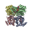
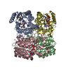
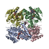
- Assembly
| Deposited unit | 
| ||||||||||||
|---|---|---|---|---|---|---|---|---|---|---|---|---|---|
| 1 | 
| ||||||||||||
| Unit cell |
| ||||||||||||
| Components on special symmetry positions |
|
-Components
| #1: Protein | Mass: 26896.027 Da / Num. of mol.: 2 Source method: isolated from a genetically manipulated source Source: (gene. exp.) Aquifex aeolicus (bacteria) / Strain: VF5 / Gene: fabG, aq_1716 / Plasmid: pET21a / Production host: Escherichia coli (E. coli) / Strain (production host): BL21-CodonPlus(DE3)-RILReferences: UniProt: O67610, 3-oxoacyl-[acyl-carrier-protein] reductase #2: Water | ChemComp-HOH / | Water Mass: 18.015 Da / Num. of mol.: 511 / Source method: isolated from a natural source / Formula: H2O Mass: 18.015 Da / Num. of mol.: 511 / Source method: isolated from a natural source / Formula: H2O |
|---|
-Experimental details
-Experiment
| Experiment | Method: X-RAY DIFFRACTION / Number of used crystals: 1 |
|---|
- Sample preparation
| Crystal | Density Matthews: 2.32 Å3/Da / Density % sol: 46.9 % |
|---|---|
| Crystal grow | Temperature: 293 K / Method: vapor diffusion, sitting drop / pH: 6.4 Details: USING 4 MICROLITER DROPS CONTAINING EQUAL VOLUMES OF PROTEIN CONCENTRATE (11.8 mg/ml) AND RESERVOIR SOLUTION CONTAINING 45% v/v PEG200, 0.1M Tris-HCl (pH 6.4), VAPOR DIFFUSION, SITTING DROP, temperature 293K |
-Data collection
| Diffraction | Mean temperature: 100 K | |||||||||||||||||||||||||||||||||||||||||||||||||||||||||||||||||||||||||||||
|---|---|---|---|---|---|---|---|---|---|---|---|---|---|---|---|---|---|---|---|---|---|---|---|---|---|---|---|---|---|---|---|---|---|---|---|---|---|---|---|---|---|---|---|---|---|---|---|---|---|---|---|---|---|---|---|---|---|---|---|---|---|---|---|---|---|---|---|---|---|---|---|---|---|---|---|---|---|---|
| Diffraction source | Source: SYNCHROTRON / Site: APS  / Beamline: 22-ID / Wavelength: 0.97243 Å / Beamline: 22-ID / Wavelength: 0.97243 Å | |||||||||||||||||||||||||||||||||||||||||||||||||||||||||||||||||||||||||||||
| Detector | Type: MARMOSAIC 300 mm CCD / Detector: CCD / Date: Oct 11, 2006 / Details: ROSENBAUM | |||||||||||||||||||||||||||||||||||||||||||||||||||||||||||||||||||||||||||||
| Radiation | Monochromator: SI CHANNEL 220 / Protocol: SINGLE WAVELENGTH / Monochromatic (M) / Laue (L): M / Scattering type: x-ray | |||||||||||||||||||||||||||||||||||||||||||||||||||||||||||||||||||||||||||||
| Radiation wavelength | Wavelength: 0.97243 Å / Relative weight: 1 | |||||||||||||||||||||||||||||||||||||||||||||||||||||||||||||||||||||||||||||
| Reflection | Redundancy: 23.7 % / Av σ(I) over netI: 20.3 / Number: 1034615 / Rmerge(I) obs: 0.089 / Χ2: 3.02 / D res high: 1.84 Å / D res low: 50 Å / Num. obs: 43626 / % possible obs: 99.7 | |||||||||||||||||||||||||||||||||||||||||||||||||||||||||||||||||||||||||||||
| Diffraction reflection shell |
| |||||||||||||||||||||||||||||||||||||||||||||||||||||||||||||||||||||||||||||
| Reflection | Resolution: 1.84→50 Å / Num. all: 43626 / Num. obs: 43626 / % possible obs: 99.7 % / Observed criterion σ(I): 0 / Redundancy: 23.7 % / Rsym value: 0.089 / Χ2: 3.016 / Net I/σ(I): 20.3 | |||||||||||||||||||||||||||||||||||||||||||||||||||||||||||||||||||||||||||||
| Reflection shell |
|
- Processing
| Software |
| ||||||||||||||||||||||||||||||||||||||||||||||||||||||||||||||||||||||||||||||||||||||||||
|---|---|---|---|---|---|---|---|---|---|---|---|---|---|---|---|---|---|---|---|---|---|---|---|---|---|---|---|---|---|---|---|---|---|---|---|---|---|---|---|---|---|---|---|---|---|---|---|---|---|---|---|---|---|---|---|---|---|---|---|---|---|---|---|---|---|---|---|---|---|---|---|---|---|---|---|---|---|---|---|---|---|---|---|---|---|---|---|---|---|---|---|
| Refinement | Method to determine structure: MOLECULAR REPLACEMENT Starting model: PDB ENTRY 2C07 Resolution: 1.84→30 Å / Cor.coef. Fo:Fc: 0.95 / Cor.coef. Fo:Fc free: 0.929 / SU B: 2.484 / SU ML: 0.079 / Cross valid method: THROUGHOUT / σ(F): 0 / ESU R: 0.142 / ESU R Free: 0.133 / Stereochemistry target values: MAXIMUM LIKELIHOOD / Details: HYDROGENS HAVE BEEN ADDED IN THE RIDING POSITIONS
| ||||||||||||||||||||||||||||||||||||||||||||||||||||||||||||||||||||||||||||||||||||||||||
| Solvent computation | Ion probe radii: 0.8 Å / Shrinkage radii: 0.8 Å / VDW probe radii: 1.2 Å / Solvent model: MASK | ||||||||||||||||||||||||||||||||||||||||||||||||||||||||||||||||||||||||||||||||||||||||||
| Displacement parameters | Biso mean: 19.301 Å2
| ||||||||||||||||||||||||||||||||||||||||||||||||||||||||||||||||||||||||||||||||||||||||||
| Refinement step | Cycle: LAST / Resolution: 1.84→30 Å
| ||||||||||||||||||||||||||||||||||||||||||||||||||||||||||||||||||||||||||||||||||||||||||
| Refine LS restraints |
| ||||||||||||||||||||||||||||||||||||||||||||||||||||||||||||||||||||||||||||||||||||||||||
| LS refinement shell | Resolution: 1.84→1.891 Å / Total num. of bins used: 20
|
