[English] 日本語
Yorodumi
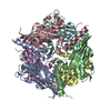
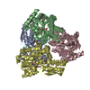
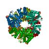
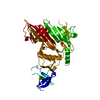
- PDB-2p5z: The E. coli c3393 protein is a component of the type VI secretion... -

+
Open data


ID or keywords:

Loading...

-
Basic information

Entry
Database: PDB / ID: 2p5z
TitleThe E. coli c3393 protein is a component of the type VI secretion system and exhibits structural similarity to T4 bacteriophage tail proteins gp27 and gp5
ComponentsType VI secretion system componentType VI secretion system
KeywordsSTRUCTURAL GENOMICS / UNKNOWN FUNCTION / PSI-2 / Protein Structure Initiative / New York SGX Research Center for Structural Genomics / NYSGXRC
Function / homology
Function and homology information


identical protein binding
Similarity search - Function
Black beetle virus capsid protein - #10 / Black beetle virus capsid protein / Pnp Oxidase; Chain A - #50 / Gp5 N-terminal domain / Baseplate protein-like domains / Type VI secretion system spike protein VgrG2, C-terminal domain of unknown function DUF2345 / Putative type VI secretion system, Rhs element associated Vgr domain / Uncharacterized protein conserved in bacteria (DUF2345) / Putative type VI secretion system Rhs element Vgr / Type VI secretion system, RhsGE-associated Vgr family subset ...Black beetle virus capsid protein - #10 / Black beetle virus capsid protein / Pnp Oxidase; Chain A - #50 / Gp5 N-terminal domain / Baseplate protein-like domains / Type VI secretion system spike protein VgrG2, C-terminal domain of unknown function DUF2345 / Putative type VI secretion system, Rhs element associated Vgr domain / Uncharacterized protein conserved in bacteria (DUF2345) / Putative type VI secretion system Rhs element Vgr / Type VI secretion system, RhsGE-associated Vgr family subset / Phage tail baseplate hub (GPD) / Phage tail protein beta-alpha-beta fold / Type VI secretion system, RhsGE-associated Vgr protein / Gp5/Type VI secretion system Vgr protein, OB-fold domain / Type VI secretion system/phage-baseplate injector OB domain / Vgr protein, OB-fold domain superfamily / 3-Layer(bab) Sandwich / Pnp Oxidase; Chain A / Few Secondary Structures / Irregular / OB fold (Dihydrolipoamide Acetyltransferase, E2P) / Roll / Beta Barrel / Mainly Beta / Alpha Beta
Similarity search - Domain/homology
Uncharacterized protein / Uncharacterized protein
Similarity search - Component
Biological speciesEscherichia coli O6 (bacteria)
MethodX-RAY DIFFRACTION / SYNCHROTRON / SAD / Resolution: 2.6 Å
AuthorsRamagopal, U.A. / Bonanno, J.B. / Sridhar, V. / Lau, C. / Toro, R. / Gheyi, T. / Maletic, M. / Freeman, J.C. / Sauder, J.M. / Burley, S.K. ...Ramagopal, U.A. / Bonanno, J.B. / Sridhar, V. / Lau, C. / Toro, R. / Gheyi, T. / Maletic, M. / Freeman, J.C. / Sauder, J.M. / Burley, S.K. / Almo, S.C. / New York SGX Research Center for Structural Genomics (NYSGXRC)
CitationJournal: Proc.Natl.Acad.Sci.Usa / Year: 2009
Title: Type VI secretion apparatus and phage tail-associated protein complexes share a common evolutionary origin.
Authors: Leiman, P.G. / Basler, M. / Ramagopal, U.A. / Bonanno, J.B. / Sauder, J.M. / Pukatzki, S. / Burley, S.K. / Almo, S.C. / Mekalanos, J.J.
History
DepositionMar 16, 2007Deposition site: RCSB / Processing site: RCSB
Revision 1.0Apr 3, 2007Provider: repository / Type: Initial release
Revision 1.1May 1, 2008Group: Version format compliance
Revision 1.2Jul 13, 2011Group: Derived calculations / Version format compliance
Revision 1.3Oct 18, 2017Group: Refinement description / Category: software
Item: _software.classification / _software.contact_author ..._software.classification / _software.contact_author / _software.contact_author_email / _software.date / _software.language / _software.location / _software.name / _software.type / _software.version
Revision 1.4Nov 14, 2018Group: Data collection / Structure summary / Category: audit_author / Item: _audit_author.identifier_ORCID
Revision 1.5Feb 3, 2021Group: Database references / Structure summary
Category: audit_author / citation_author / struct_ref_seq_dif
Item: _audit_author.identifier_ORCID / _citation_author.identifier_ORCID / _struct_ref_seq_dif.details
Revision 1.6Feb 21, 2024Group: Data collection / Database references / Category: chem_comp_atom / chem_comp_bond / database_2
Item: _database_2.pdbx_DOI / _database_2.pdbx_database_accession

-
Structure visualization

Structure viewerMolecule:
MolmilJmol/JSmol

Downloads & links

-
Assembly

Deposited unit
X: Type VI secretion system component


Theoretical massNumber of molelcules
Total (without water)55,6981
Polymers55,6981
Non-polymers00
Water1,11762
1
X: Type VI secretion system component

X: Type VI secretion system component

X: Type VI secretion system component


Theoretical massNumber of molelcules
Total (without water)167,0943
Polymers167,0943
Non-polymers00
Water543
TypeNameSymmetry operationNumber
identity operation1_555x,y,z1
crystal symmetry operation2_655-y+1,x-y,z1
crystal symmetry operation3_665-x+y+1,-x+1,z1
Buried area4440 Å2
ΔGint-21 kcal/mol
Surface area50070 Å2
MethodPISA, PQS
Unit cell
Length a, b, c (Å)116.351, 116.351, 80.584
Angle α, β, γ (deg.)90.000, 90.000, 120.000
Int Tables number173
Space group name H-MP63
DetailsBiological unit appears to be trimer

-
Components

#1: Protein Type VI secretion system component / Type VI secretion system


Mass: 55697.988 Da / Num. of mol.: 1 / Fragment: N-terminal domain
Source method: isolated from a genetically manipulated source
Source: (gene. exp.) Escherichia coli O6 (bacteria) / Species: Escherichia coli / Strain: O6:H1, CFT073, UPEC / Gene: c3393 / Plasmid: BC-pSGX3(BC) / Species (production host): Escherichia coli / Production host: Escherichia coli BL21(DE3) (bacteria) / Strain (production host): BL21(DE3) / References: UniProt: Q8FED2, UniProt: A0A0H2VBP9*PLUS
#2: Water ChemComp-HOH / water / Water


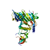
Mass: 18.015 Da / Num. of mol.: 62 / Source method: isolated from a natural source / Formula: H2O

-
Experimental details

-
Experiment

ExperimentMethod: X-RAY DIFFRACTION / Number of used crystals: 1

-
Sample preparation

CrystalDensity Matthews: 2.83 Å3/Da / Density % sol: 56.47 %
Crystal growTemperature: 298 K / Method: vapor diffusion, sitting drop / pH: 6.5
Details: 0.1M Hepes pH 7.5, 21% PEG 3350, pH 6.5, VAPOR DIFFUSION, SITTING DROP, temperature 298K

-
Data collection

DiffractionMean temperature: 100 K
Diffraction sourceSource: SYNCHROTRON / Site: APS / Beamline: 31-ID / Wavelength: 0.9793 Å
DetectorType: MAR CCD 165 mm / Detector: CCD / Date: Feb 12, 2007
RadiationProtocol: SINGLE WAVELENGTH / Monochromatic (M) / Laue (L): M / Scattering type: x-ray
Radiation wavelengthWavelength: 0.9793 Å / Relative weight: 1
ReflectionResolution: 2.6→50 Å / Num. all: 19128 / Num. obs: 19128 / % possible obs: 100 % / Observed criterion σ(F): 0 / Observed criterion σ(I): 0 / Redundancy: 15.5 % / Rmerge(I) obs: 0.107 / Rsym value: 0.095 / Χ2: 1.442 / Net I/σ(I): 6.6
Reflection shellResolution: 2.6→2.69 Å / Redundancy: 15.7 % / Rmerge(I) obs: 0.753 / Mean I/σ(I) obs: 2.8 / Num. unique all: 1892 / Rsym value: 0.558 / Χ2: 0.427 / % possible all: 100

-
Processing

Software
NameVersionClassificationNB
DENZOdata reduction
SCALEPACKdata scaling
REFMACrefinement
PDB_EXTRACT2data extraction
MAR345CCDdata collection
HKL-2000data reduction
SHELXDphasing
SHELXEmodel building
RefinementMethod to determine structure: SAD / Resolution: 2.6→47.19 Å / Cor.coef. Fo:Fc: 0.94 / Cor.coef. Fo:Fc free: 0.926 / SU B: 10.191 / SU ML: 0.218 / Cross valid method: THROUGHOUT / σ(F): 0 / σ(I): 0 / ESU R: 0.395 / ESU R Free: 0.283 / Stereochemistry target values: MAXIMUM LIKELIHOOD / Details: HYDROGENS HAVE BEEN ADDED IN THE RIDING POSITIONS
RfactorNum. reflection% reflectionSelection details
Rfree0.264 983 5.1 %RANDOM
Rwork0.217 ---
all0.219 19100 --
obs0.219 19100 99.9 %-
Solvent computationIon probe radii: 0.8 Å / Shrinkage radii: 0.8 Å / VDW probe radii: 1.2 Å / Solvent model: MASK
Displacement parametersBiso mean: 60.297 Å2
Baniso -1Baniso -2Baniso -3
1-1.57 Å20.78 Å20 Å2
2--1.57 Å20 Å2
3----2.35 Å2
Refinement stepCycle: LAST / Resolution: 2.6→47.19 Å
ProteinNucleic acidLigandSolventTotal
Num. atoms2891 0 0 62 2953
Refine LS restraints
Refine-IDTypeDev idealDev ideal targetNumber
X-RAY DIFFRACTIONr_bond_refined_d0.0110.0222959
X-RAY DIFFRACTIONr_angle_refined_deg1.8741.9624001
X-RAY DIFFRACTIONr_dihedral_angle_1_deg8.5155356
X-RAY DIFFRACTIONr_dihedral_angle_2_deg34.55322.69145
X-RAY DIFFRACTIONr_dihedral_angle_3_deg20.27115502
X-RAY DIFFRACTIONr_dihedral_angle_4_deg17.1541531
X-RAY DIFFRACTIONr_chiral_restr0.1410.2439
X-RAY DIFFRACTIONr_gen_planes_refined0.0060.022259
X-RAY DIFFRACTIONr_nbd_refined0.160.11203
X-RAY DIFFRACTIONr_nbtor_refined0.3220.31957
X-RAY DIFFRACTIONr_xyhbond_nbd_refined0.2110.5203
X-RAY DIFFRACTIONr_symmetry_vdw_refined0.1110.138
X-RAY DIFFRACTIONr_symmetry_hbond_refined0.1820.57
X-RAY DIFFRACTIONr_mcbond_it2.89621832
X-RAY DIFFRACTIONr_mcangle_it4.63432894
X-RAY DIFFRACTIONr_scbond_it3.10521263
X-RAY DIFFRACTIONr_scangle_it4.65831107
LS refinement shellResolution: 2.6→2.673 Å / Total num. of bins used: 20
RfactorNum. reflection% reflection
Rfree0.457 89 -
Rwork0.318 1321 -
obs-1410 99.86 %

+
About Yorodumi

-
News

-
Feb 9, 2022. New format data for meta-information of EMDB entries

New format data for meta-information of EMDB entries

  • Version 3 of the EMDB header file is now the official format.
  • The previous official version 1.9 will be removed from the archive.

Related info.:EMDB header

External links:wwPDB to switch to version 3 of the EMDB data model

-
Aug 12, 2020. Covid-19 info

Covid-19 info

URL: https://pdbj.org/emnavi/covid19.php

New page: Covid-19 featured information page in EM Navigator.

Related info.:Covid-19 info / Mar 5, 2020. Novel coronavirus structure data

+
Mar 5, 2020. Novel coronavirus structure data

Novel coronavirus structure data

Related info.:Yorodumi Speices / Aug 12, 2020. Covid-19 info

External links:COVID-19 featured content - PDBj / Molecule of the Month (242):Coronavirus Proteases

+
Jan 31, 2019. EMDB accession codes are about to change! (news from PDBe EMDB page)

EMDB accession codes are about to change! (news from PDBe EMDB page)

  • The allocation of 4 digits for EMDB accession codes will soon come to an end. Whilst these codes will remain in use, new EMDB accession codes will include an additional digit and will expand incrementally as the available range of codes is exhausted. The current 4-digit format prefixed with “EMD-” (i.e. EMD-XXXX) will advance to a 5-digit format (i.e. EMD-XXXXX), and so on. It is currently estimated that the 4-digit codes will be depleted around Spring 2019, at which point the 5-digit format will come into force.
  • The EM Navigator/Yorodumi systems omit the EMD- prefix.

Related info.:Q: What is EMD? / ID/Accession-code notation in Yorodumi/EM Navigator

External links:EMDB Accession Codes are Changing Soon! / Contact to PDBj

+
Jul 12, 2017. Major update of PDB

Major update of PDB

  • wwPDB released updated PDB data conforming to the new PDBx/mmCIF dictionary.
  • This is a major update changing the version number from 4 to 5, and with Remediation, in which all the entries are updated.
  • In this update, many items about electron microscopy experimental information are reorganized (e.g. em_software).
  • Now, EM Navigator and Yorodumi are based on the updated data.

External links:wwPDB Remediation / Enriched Model Files Conforming to OneDep Data Standards Now Available in the PDB FTP Archive

-
Yorodumi

Thousand views of thousand structures

  • Yorodumi is a browser for structure data from EMDB, PDB, SASBDB, etc.
  • This page is also the successor to EM Navigator detail page, and also detail information page/front-end page for Omokage search.
  • The word "yorodu" (or yorozu) is an old Japanese word meaning "ten thousand". "mi" (miru) is to see.

Related info.:EMDB / PDB / SASBDB / Comparison of 3 databanks / Yorodumi Search / Aug 31, 2016. New EM Navigator & Yorodumi / Yorodumi Papers / Jmol/JSmol / Function and homology information / Changes in new EM Navigator and Yorodumi

Read more