 Movie
Movie Controller

[English] 日本語
 Yorodumi
- PDB-2p4g: CRYSTAL STRUCTURE OF A PYRIMIDINE REDUCTASE-LIKE PROTEIN (DIP1392... -
+ Open data

- Basic information
| Entry | Database: PDB / ID: 2p4g | ||||||
|---|---|---|---|---|---|---|---|
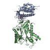
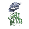
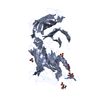
| Title | CRYSTAL STRUCTURE OF A PYRIMIDINE REDUCTASE-LIKE PROTEIN (DIP1392) FROM CORYNEBACTERIUM DIPHTHERIAE NCTC AT 2.30 A RESOLUTION | ||||||
 Components Components | Hypothetical protein | ||||||
 Keywords Keywords | OXIDOREDUCTASE / PYRIMIDINE REDUCTASE-LIKE PROTEIN / STRUCTURAL GENOMICS / JOINT CENTER FOR STRUCTURAL GENOMICS / JCSG / PROTEIN STRUCTURE INITIATIVE / PSI-2 | ||||||
| Function / homology |  Function and homology information5-amino-6-(5-phosphoribosylamino)uracil reductase activity / riboflavin biosynthetic process Function and homology information5-amino-6-(5-phosphoribosylamino)uracil reductase activity / riboflavin biosynthetic processSimilarity search - Function | ||||||
| Biological species |  Corynebacterium diphtheriae (bacteria) Corynebacterium diphtheriae (bacteria) | ||||||
| Method | X-RAY DIFFRACTION / SYNCHROTRON / MAD / Resolution: 2.3 Å | ||||||
 Authors Authors | Joint Center for Structural Genomics (JCSG) | ||||||
 Citation Citation | Journal: To be published Title: Crystal structure of hypothetical protein (NP_939744.1) from Corynebacterium diphtheriae at 2.30 A resolution Authors: Joint Center for Structural Genomics (JCSG) | ||||||
| History |
| ||||||
| Remark 300 | BIOMOLECULE: 1 THIS ENTRY CONTAINS THE CRYSTALLOGRAPHIC ASYMMETRIC UNIT WHICH CONSISTS OF 1 ... BIOMOLECULE: 1 THIS ENTRY CONTAINS THE CRYSTALLOGRAPHIC ASYMMETRIC UNIT WHICH CONSISTS OF 1 CHAIN(S). SEE REMARK 350 FOR INFORMATION ON GENERATING THE BIOLOGICAL MOLECULE(S). SIZE EXCLUSION CHROMATOGRAPHY SUPPORTS THE ASSIGNMENT OF A DIMER AS A BIOLOGICALLY SIGNIFICANT OLIGOMERIZATION STATE. | ||||||
| Remark 999 | SEQUENCE THE CONSTRUCT WAS EXPRESSED WITH A PURIFICATION TAG MGSDKIHHHHHHENLYFQG. THE TAG WAS ... SEQUENCE THE CONSTRUCT WAS EXPRESSED WITH A PURIFICATION TAG MGSDKIHHHHHHENLYFQG. THE TAG WAS REMOVED WITH TEV PROTEASE LEAVING ONLY A GLYCINE (0), FOLLOWED BY THE TARGET SEQUENCE. |
- Structure visualization
| Structure viewer | Molecule: MolmilJmol/JSmol |
|---|
- Downloads & links
-Download
| PDBx/mmCIF format | 2p4g.cif.gz | 63.9 KB | Display | PDBx/mmCIF format |
|---|---|---|---|---|
| PDB format | pdb2p4g.ent.gz | 49 KB | Display | PDB format |
| PDBx/mmJSON format | 2p4g.json.gz | Tree view | PDBx/mmJSON format | |
| Others |  Other downloads Other downloads |
-Validation report
| Arichive directory | https://data.pdbj.org/pub/pdb/validation_reports/p4/2p4gftp://data.pdbj.org/pub/pdb/validation_reports/p4/2p4g | HTTPS FTP |
|---|
-Related structure data
| Similar structure data | |
|---|---|
| Other databases |


-Links
 PDBj
PDBj
- Assembly
| Deposited unit | 
| ||||||||
|---|---|---|---|---|---|---|---|---|---|
| 1 | 
| ||||||||
| Unit cell |
| ||||||||
| Details | SIZE EXCLUSION CHROMATOGRAPHY SUPPORTS THE ASSIGNMENT OF A DIMER AS A BIOLOGICALLY SIGNIFICANT OLIGOMERIZATION STATE. |
-Components
| #1: Protein | Mass: 29485.064 Da / Num. of mol.: 1 Source method: isolated from a genetically manipulated source Source: (gene. exp.) Corynebacterium diphtheriae (bacteria) / Strain: NCTC 13129 / Gene: NP_939744.1, DIP1392 / Plasmid: speedET / Production host: Escherichia coli (E. coli) / Strain (production host): HK100 / References: UniProt: Q6NGV8 | ||
|---|---|---|---|
| #2: Chemical | ChemComp-NO3 / Nitrate  Mass: 62.005 Da / Num. of mol.: 1 / Source method: obtained synthetically / Formula: NO3 Mass: 62.005 Da / Num. of mol.: 1 / Source method: obtained synthetically / Formula: NO3 | ||
| #3: Chemical | ChemComp-EDO / Ethylene glycol  Mass: 62.068 Da / Num. of mol.: 15 / Source method: obtained synthetically / Formula: C2H6O2 Mass: 62.068 Da / Num. of mol.: 15 / Source method: obtained synthetically / Formula: C2H6O2#4: Water | ChemComp-HOH / | Water Mass: 18.015 Da / Num. of mol.: 113 / Source method: isolated from a natural source / Formula: H2O Mass: 18.015 Da / Num. of mol.: 113 / Source method: isolated from a natural source / Formula: H2O |
-Experimental details
-Experiment
| Experiment | Method: X-RAY DIFFRACTION / Number of used crystals: 2 |
|---|
- Sample preparation
| Crystal |
| |||||||||||||||
|---|---|---|---|---|---|---|---|---|---|---|---|---|---|---|---|---|
| Crystal grow |
|
-Data collection
| Diffraction |
| ||||||||||||||||||||||||||||||||||||||||||||||||||||||||||||||||||
|---|---|---|---|---|---|---|---|---|---|---|---|---|---|---|---|---|---|---|---|---|---|---|---|---|---|---|---|---|---|---|---|---|---|---|---|---|---|---|---|---|---|---|---|---|---|---|---|---|---|---|---|---|---|---|---|---|---|---|---|---|---|---|---|---|---|---|---|
| Diffraction source |
| ||||||||||||||||||||||||||||||||||||||||||||||||||||||||||||||||||
| Detector |
| ||||||||||||||||||||||||||||||||||||||||||||||||||||||||||||||||||
| Radiation |
| ||||||||||||||||||||||||||||||||||||||||||||||||||||||||||||||||||
| Radiation wavelength |
| ||||||||||||||||||||||||||||||||||||||||||||||||||||||||||||||||||
| Reflection | Resolution: 2.3→28.273 Å / Num. obs: 18260 / % possible obs: 97.3 % / Redundancy: 3.66 % / Biso Wilson estimate: 46.09 Å2 / Rmerge(I) obs: 0.052 / Net I/σ(I): 11.61 | ||||||||||||||||||||||||||||||||||||||||||||||||||||||||||||||||||
| Reflection shell |
|

-Phasing
| Phasing | Method: MAD |
|---|
- Processing
| Software |
| |||||||||||||||||||||||||||||||||||||||||||||||||||||||||||||||||||||||||||||||||||||||||||||||||||||||||||||||||||||||||||||
|---|---|---|---|---|---|---|---|---|---|---|---|---|---|---|---|---|---|---|---|---|---|---|---|---|---|---|---|---|---|---|---|---|---|---|---|---|---|---|---|---|---|---|---|---|---|---|---|---|---|---|---|---|---|---|---|---|---|---|---|---|---|---|---|---|---|---|---|---|---|---|---|---|---|---|---|---|---|---|---|---|---|---|---|---|---|---|---|---|---|---|---|---|---|---|---|---|---|---|---|---|---|---|---|---|---|---|---|---|---|---|---|---|---|---|---|---|---|---|---|---|---|---|---|---|---|---|
| Refinement | Method to determine structure: MAD / Resolution: 2.3→28.273 Å / Cor.coef. Fo:Fc: 0.963 / Cor.coef. Fo:Fc free: 0.936 / SU B: 11.84 / SU ML: 0.145 / TLS residual ADP flag: LIKELY RESIDUAL / Cross valid method: THROUGHOUT / σ(F): 0 / ESU R: 0.193 / ESU R Free: 0.183 / Stereochemistry target values: MAXIMUM LIKELIHOOD Details: 1. ATOM RECORD CONTAINS RESIDUAL B FACTORS ONLY. 2. HYDROGENS HAVE BEEN ADDED IN THE RIDING POSITIONS. 3. ANALYSIS OF THE DIFFRACTION DATA INDICATED HEMIHEDERAL TWINNING WITH A TWIN FRACTION ...Details: 1. ATOM RECORD CONTAINS RESIDUAL B FACTORS ONLY. 2. HYDROGENS HAVE BEEN ADDED IN THE RIDING POSITIONS. 3. ANALYSIS OF THE DIFFRACTION DATA INDICATED HEMIHEDERAL TWINNING WITH A TWIN FRACTION OF 0.135 AND WITH A TWIN LAW OF K,H,-L. 4. THE INITIAL REFINEMENT WAS AGAINST THE NON-DETWINNED DATA AND INCLUDED PHASE RESTRAINTS TO THE EXPERIMENTAL MAD PHASES FROM ANOTHER CRYSTAL. 5. FOR THE FINAL STAGES OF REFINEMENT, THE DIFFRACTION INTENSITIES WERE DETWINNED USING THE CCP4 PROGAM DETWIN WITH A TWIN FRACTION OF 0.135 AND A TWIN OPERATION OF K,H,-L. 6. ETHYLENE GLYCOL USED AS A CRYOPROTECTANT AND NITRATE FROM THE CRYSTALLIZATION WERE MODELED INTO THE STRUCTURE. 7. ELECTRON DENSITIES FOR MSE 1-THR 14 AND THR 263-GLN 269 WERE DISORDERED, THEREFORE THESE RESIDUES WERE NOT MODELED. 8. A MET-INHIBITION PROTOCOL WAS USED FOR SELENOMETHIONINE INCORPORATION DURING PROTEIN EXPRESSION. THE OCCUPANCY OF THE SE ATOMS IN THE MSE RESIDUES WAS REDUCED TO 0.75 TO ACCOUNT FOR THE REDUCED SCATTERING POWER DUE TO PARTIAL S-MET INCORPORATION. 9. ASP 233 IS A RAMACHANDRAN OUTLIER EVEN THOUGH IT IS MODELED INTO WELL-ORDERED ELECTRON DENSITY. 10. UNEXPLAINED NEGATIVE DIFFERENCE ELECTRON DENSITY IS OBSERVED BETWEEN LEU 203 AND LEU 211.
| |||||||||||||||||||||||||||||||||||||||||||||||||||||||||||||||||||||||||||||||||||||||||||||||||||||||||||||||||||||||||||||
| Solvent computation | Ion probe radii: 0.8 Å / Shrinkage radii: 0.8 Å / VDW probe radii: 1.2 Å / Solvent model: BABINET MODEL WITH MASK | |||||||||||||||||||||||||||||||||||||||||||||||||||||||||||||||||||||||||||||||||||||||||||||||||||||||||||||||||||||||||||||
| Displacement parameters | Biso mean: 60.7 Å2
| |||||||||||||||||||||||||||||||||||||||||||||||||||||||||||||||||||||||||||||||||||||||||||||||||||||||||||||||||||||||||||||
| Refinement step | Cycle: LAST / Resolution: 2.3→28.273 Å
| |||||||||||||||||||||||||||||||||||||||||||||||||||||||||||||||||||||||||||||||||||||||||||||||||||||||||||||||||||||||||||||
| Refine LS restraints |
| |||||||||||||||||||||||||||||||||||||||||||||||||||||||||||||||||||||||||||||||||||||||||||||||||||||||||||||||||||||||||||||
| LS refinement shell | Resolution: 2.3→2.362 Å / Total num. of bins used: 20
| |||||||||||||||||||||||||||||||||||||||||||||||||||||||||||||||||||||||||||||||||||||||||||||||||||||||||||||||||||||||||||||
| Refinement TLS params. | Method: refined / Origin x: 38.473 Å / Origin y: 32.615 Å / Origin z: 18.843 Å
| |||||||||||||||||||||||||||||||||||||||||||||||||||||||||||||||||||||||||||||||||||||||||||||||||||||||||||||||||||||||||||||
| Refinement TLS group | Selection: ALL |
