[English] 日本語
Yorodumi
- PDB-2p2s: Crystal structure of putative oxidoreductase (YP_050235.1) from E... -

+
Open data


ID or keywords:

Loading...

-
Basic information

Entry
Database: PDB / ID: 2p2s
TitleCrystal structure of putative oxidoreductase (YP_050235.1) from Erwinia carotovora atroseptica SCRI1043 at 1.25 A resolution
ComponentsPutative oxidoreductase
KeywordsOXIDOREDUCTASE / YP_050235.1 / putative / Structural Genomics / Joint Center for Structural Genomics / JCSG / Protein Structure Initiative / PSI-2
Function / homology
Function and homology information


oxidoreductase activity / metal ion binding
Similarity search - Function
Gfo/Idh/MocA-like oxidoreductase, C-terminal / Oxidoreductase family, C-terminal alpha/beta domain / Gfo/Idh/MocA-like oxidoreductase, N-terminal / Oxidoreductase family, NAD-binding Rossmann fold / Dihydrodipicolinate Reductase; domain 2 / Dihydrodipicolinate Reductase; domain 2 / NAD(P)-binding Rossmann-like Domain / NAD(P)-binding domain superfamily / Rossmann fold / 2-Layer Sandwich ...Gfo/Idh/MocA-like oxidoreductase, C-terminal / Oxidoreductase family, C-terminal alpha/beta domain / Gfo/Idh/MocA-like oxidoreductase, N-terminal / Oxidoreductase family, NAD-binding Rossmann fold / Dihydrodipicolinate Reductase; domain 2 / Dihydrodipicolinate Reductase; domain 2 / NAD(P)-binding Rossmann-like Domain / NAD(P)-binding domain superfamily / Rossmann fold / 2-Layer Sandwich / 3-Layer(aba) Sandwich / Alpha Beta
Similarity search - Domain/homology
ACETATE ION / Putative oxidoreductase
Similarity search - Component
Biological speciesPectobacterium atrosepticum SCRI1043 (bacteria)
MethodX-RAY DIFFRACTION / SYNCHROTRON / MAD / Resolution: 1.25 Å
AuthorsJoint Center for Structural Genomics (JCSG)
CitationJournal: To be Published
Title: Crystal structure of putative oxidoreductase (YP_050235.1) from Erwinia carotovora atroseptica SCRI1043 at 1.25 A resolution
Authors: Joint Center for Structural Genomics (JCSG)
History
DepositionMar 7, 2007Deposition site: RCSB / Processing site: RCSB
Revision 1.0Mar 20, 2007Provider: repository / Type: Initial release
Revision 1.1May 1, 2008Group: Version format compliance
Revision 1.2Jul 13, 2011Group: Advisory / Version format compliance
Revision 1.3Oct 18, 2017Group: Refinement description / Category: software / Item: _software.classification / _software.name
Revision 1.4Oct 25, 2017Group: Author supporting evidence / Category: pdbx_struct_assembly_auth_evidence
Revision 1.5Jan 25, 2023Group: Database references / Derived calculations
Category: database_2 / pdbx_struct_conn_angle ...database_2 / pdbx_struct_conn_angle / struct_conn / struct_ref_seq_dif / struct_site
Item: _database_2.pdbx_DOI / _database_2.pdbx_database_accession ..._database_2.pdbx_DOI / _database_2.pdbx_database_accession / _pdbx_struct_conn_angle.ptnr1_auth_asym_id / _pdbx_struct_conn_angle.ptnr1_auth_comp_id / _pdbx_struct_conn_angle.ptnr1_auth_seq_id / _pdbx_struct_conn_angle.ptnr1_label_asym_id / _pdbx_struct_conn_angle.ptnr1_label_atom_id / _pdbx_struct_conn_angle.ptnr1_label_comp_id / _pdbx_struct_conn_angle.ptnr1_label_seq_id / _pdbx_struct_conn_angle.ptnr1_symmetry / _pdbx_struct_conn_angle.ptnr3_auth_asym_id / _pdbx_struct_conn_angle.ptnr3_auth_comp_id / _pdbx_struct_conn_angle.ptnr3_auth_seq_id / _pdbx_struct_conn_angle.ptnr3_label_asym_id / _pdbx_struct_conn_angle.ptnr3_label_atom_id / _pdbx_struct_conn_angle.ptnr3_label_comp_id / _pdbx_struct_conn_angle.ptnr3_label_seq_id / _pdbx_struct_conn_angle.ptnr3_symmetry / _pdbx_struct_conn_angle.value / _struct_conn.conn_type_id / _struct_conn.id / _struct_conn.pdbx_dist_value / _struct_conn.pdbx_leaving_atom_flag / _struct_conn.ptnr1_auth_asym_id / _struct_conn.ptnr1_auth_comp_id / _struct_conn.ptnr1_auth_seq_id / _struct_conn.ptnr1_label_asym_id / _struct_conn.ptnr1_label_atom_id / _struct_conn.ptnr1_label_comp_id / _struct_conn.ptnr1_label_seq_id / _struct_conn.ptnr1_symmetry / _struct_conn.ptnr2_auth_asym_id / _struct_conn.ptnr2_auth_comp_id / _struct_conn.ptnr2_auth_seq_id / _struct_conn.ptnr2_label_asym_id / _struct_conn.ptnr2_label_atom_id / _struct_conn.ptnr2_label_comp_id / _struct_conn.ptnr2_label_seq_id / _struct_conn.ptnr2_symmetry / _struct_ref_seq_dif.details / _struct_site.pdbx_auth_asym_id / _struct_site.pdbx_auth_comp_id / _struct_site.pdbx_auth_seq_id
Remark 300 
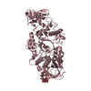
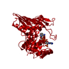
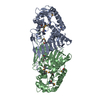
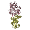
BIOMOLECULE: 1 THIS ENTRY CONTAINS THE CRYSTALLOGRAPHIC ASYMMETRIC UNIT WHICH CONSISTS OF 2 ... BIOMOLECULE: 1 THIS ENTRY CONTAINS THE CRYSTALLOGRAPHIC ASYMMETRIC UNIT WHICH CONSISTS OF 2 CHAIN(S). SEE REMARK 350 FOR INFORMATION ON GENERATING THE BIOLOGICAL MOLECULE(S). SIZE EXCLUSION CHROMATOGRAPHY WITH STATIC LIGHT SCATTERING SUPPORTS THE ASSIGNMENT OF A DIMER AS A BIOLOGICALLY SIGNIFICANT OLIGOMERIZATION STATE.
Remark 999 SEQUENCE THE CONSTRUCT WAS EXPRESSED WITH A PURIFICATION TAG MGSDKIHHHHHHENLYFQG. THE TAG WAS ... SEQUENCE THE CONSTRUCT WAS EXPRESSED WITH A PURIFICATION TAG MGSDKIHHHHHHENLYFQG. THE TAG WAS REMOVED WITH TEV PROTEASE LEAVING ONLY A GLYCINE (0) FOLLOWED BY THE TARGET SEQUENCE.

-
Structure visualization

Structure viewerMolecule:
MolmilJmol/JSmol

Downloads & links

-
Assembly

Deposited unit
A: Putative oxidoreductase
B: Putative oxidoreductase
hetero molecules


Theoretical massNumber of molelcules
Total (without water)75,89311
Polymers75,4132
Non-polymers4809
Water15,295849
1


  • Idetical with deposited unit
  • defined by author&software
  • Evidence: gel filtration, light scattering
TypeNameSymmetry operationNumber
identity operation1_555x,y,z1
Buried area5780 Å2
ΔGint-17 kcal/mol
Surface area24560 Å2
MethodPISA
Unit cell
Length a, b, c (Å)188.050, 45.830, 87.970
Angle α, β, γ (deg.)90.000, 116.400, 90.000
Int Tables number5
Space group name H-MC121
Components on special symmetry positions
IDModelComponents
11B-688-

HOH

Noncrystallographic symmetry (NCS)NCS domain:
IDEns-IDDetails
11A
21B

NCS domain segments:

Component-ID: 1 / Ens-ID: 1 / Beg label comp-ID: LYS / End label comp-ID: ALA / Refine code: 5 / Auth seq-ID: 2 - 335 / Label seq-ID: 3 - 336

Dom-IDAuth asym-IDLabel asym-ID
1AA
2BB
DetailsSIZE EXCLUSION CHROMATOGRAPHY WITH STATIC LIGHT SCATTERING SUPPORTS THE ASSIGNMENT OF A DIMER AS A BIOLOGICALLY SIGNIFICANT OLIGOMERIZATION STATE.

-
Components

#1: Protein Putative oxidoreductase /


Mass: 37706.613 Da / Num. of mol.: 2
Source method: isolated from a genetically manipulated source
Source: (gene. exp.) Pectobacterium atrosepticum SCRI1043 (bacteria)
Species: Pectobacterium atrosepticum / Strain: SCRI 1043 / Gene: YP_050235.1, ECA2140 / Plasmid: speedET / Production host: Escherichia coli (E. coli) / Strain (production host): HK100
References: UniProt: Q6D5A0, Oxidoreductases; Acting on the CH-OH group of donors
#2: Chemical ChemComp-MG / MAGNESIUM ION


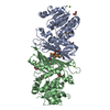
Mass: 24.305 Da / Num. of mol.: 2 / Source method: obtained synthetically / Formula: Mg
#3: Chemical
ChemComp-EDO / 1,2-ETHANEDIOL / ETHYLENE GLYCOL / Ethylene glycol


Mass: 62.068 Da / Num. of mol.: 6 / Source method: obtained synthetically / Formula: C2H6O2
#4: Chemical ChemComp-ACT / ACETATE ION / Acetate


Mass: 59.044 Da / Num. of mol.: 1 / Source method: obtained synthetically / Formula: C2H3O2
#5: Water ChemComp-HOH / water / Water


Mass: 18.015 Da / Num. of mol.: 849 / Source method: isolated from a natural source / Formula: H2O

-
Experimental details

-
Experiment

ExperimentMethod: X-RAY DIFFRACTION / Number of used crystals: 1

-
Sample preparation

Crystal
IDDensity Matthews3/Da)Density % sol (%)Description
12.2545.34THE STRUCTURE WAS SOLVED BY MAD METHOD USING A DIFFERENT CRYSTAL. THE PHASE RESTRAINTS FROM THAT CRYSTAL WERE USED IN THE CURRENT REFINEMENT
2THE STRUCTURE WAS SOLVED BY MAD METHOD USING A DIFFERENT CRYSTAL. THE PHASE RESTRAINTS FROM THAT CRYSTAL WERE USED IN THE CURRENT REFINEMENT
3THE STRUCTURE WAS SOLVED BY MAD METHOD USING A DIFFERENT CRYSTAL. THE PHASE RESTRAINTS FROM THAT CRYSTAL WERE USED IN THE CURRENT REFINEMENT
Crystal grow
Temperature (K)Crystal-IDMethodpHDetails
2931vapor diffusion, sitting drop6.33NANODROP, 20.0% PEG 8000, 0.2M Magnesium acetate, 0.1M Sodium cacodylate pH 6.33, VAPOR DIFFUSION, SITTING DROP, temperature 293K
2932vapor diffusion, sitting drop7NANODROP, 22.0% PEG 8000, 0.15M Magnesium acetate, 0.1M Sodium cacodylate pH 7.0, VAPOR DIFFUSION, SITTING DROP, temperature 293K

-
Data collection

Diffraction
IDMean temperature (K)Crystal-ID
11001
21002
Diffraction source
SourceSiteBeamlineIDWavelength (Å)
SYNCHROTRONSSRL BL11-111
SYNCHROTRONSSRL BL9-220.91837, 0.97945, 0.97927
Detector
TypeIDDetectorDateDetails
MARMOSAIC 325 mm CCD1CCDFeb 24, 2007Flat mirror (vertical focusing)
MARMOSAIC 325 mm CCD2CCDFeb 16, 2007Flat collimating mirror, toroid focusing mirror
Radiation
IDMonochromatorProtocolMonochromatic (M) / Laue (L)Scattering typeWavelength-ID
1Double crystalSINGLE WAVELENGTHMx-ray1
2Double crystalMADMx-ray1
Radiation wavelength
IDWavelength (Å)Relative weight
111
20.918371
30.979451
40.979271
ReflectionResolution: 1.25→47.891 Å / Num. obs: 184625 / % possible obs: 95.4 % / Biso Wilson estimate: 16.288 Å2 / Rmerge(I) obs: 0.079 / Net I/σ(I): 12.56
Reflection shell

Diffraction-ID: 1,2

Resolution (Å)Highest resolution (Å)Rmerge(I) obsMean I/σ(I) obsNum. measured obsNum. unique all% possible all
1.25-1.30.5032.4545621835489.3
1.3-1.360.4112.9569531901291.4
1.36-1.430.3353.6565791880093.5
1.43-1.520.2434.9593921965395.1
1.52-1.640.1617.2617942034596.8
1.64-1.810.10410.6627052054697.9
1.81-2.070.08517.3669021996698.1
2.070.10826.11148112037498.8

-
Phasing

PhasingMethod: MAD

-
Processing

Software
NameVersionClassificationNB
MolProbity3beta29model building
SHELXphasing
REFMAC5.2.0019refinement
XSCALEdata scaling
PDB_EXTRACT2data extraction
MAR345CCDdata collection
XDSdata reduction
SHELXDphasing
autoSHARPphasing
RefinementMethod to determine structure: MAD / Resolution: 1.25→47.891 Å / Cor.coef. Fo:Fc: 0.975 / Cor.coef. Fo:Fc free: 0.967 / SU B: 1.359 / SU ML: 0.029 / TLS residual ADP flag: LIKELY RESIDUAL / Cross valid method: THROUGHOUT / σ(F): 0 / ESU R: 0.041 / ESU R Free: 0.043
Stereochemistry target values: MAXIMUM LIKELIHOOD WITH PHASES
Details: 1. HYDROGENS HAVE BEEN ADDED IN THE RIDING POSITIONS 2. ATOM RECORD CONTAINS RESIDUAL B FACTORS ONLY. 3. A MET-INHIBITION PROTOCOL WAS USED FOR SELENOMETHIONINE INCORPORATION DURING PROTEIN ...Details: 1. HYDROGENS HAVE BEEN ADDED IN THE RIDING POSITIONS 2. ATOM RECORD CONTAINS RESIDUAL B FACTORS ONLY. 3. A MET-INHIBITION PROTOCOL WAS USED FOR SELENOMETHIONINE INCORPORATION DURING PROTEIN EXPRESSION. THE OCCUPANCY OF THE SE ATOMS IN THE MSE RESIDUES WAS REDUCED TO 0.75 FOR THE REDUCED SCATTERING POWER DUE TO PARTIAL S-MET INCORPORATION. 4. ETHYLENE GLYCOL, ACETATE AND MAGNESIUM IONS ARE MODELED BASED ON CRYSTALLIZATION AND CRYO CONDITIONS.
RfactorNum. reflection% reflectionSelection details
Rfree0.179 9253 5 %RANDOM
Rwork0.156 ---
all0.157 ---
obs0.157 184625 99.35 %-
Solvent computationIon probe radii: 0.8 Å / Shrinkage radii: 0.8 Å / VDW probe radii: 1.2 Å / Solvent model: BABINET MODEL WITH MASK
Displacement parametersBiso mean: 9.215 Å2
Baniso -1Baniso -2Baniso -3
1--0.13 Å20 Å2-0.12 Å2
2--0.22 Å20 Å2
3----0.2 Å2
Refinement stepCycle: LAST / Resolution: 1.25→47.891 Å
ProteinNucleic acidLigandSolventTotal
Num. atoms5221 0 30 849 6100
Refine LS restraints
Refine-IDTypeDev idealDev ideal targetNumber
X-RAY DIFFRACTIONr_bond_refined_d0.0150.0225690
X-RAY DIFFRACTIONr_bond_other_d0.0010.023919
X-RAY DIFFRACTIONr_angle_refined_deg1.6171.9427763
X-RAY DIFFRACTIONr_angle_other_deg0.97339505
X-RAY DIFFRACTIONr_dihedral_angle_1_deg6.0345759
X-RAY DIFFRACTIONr_dihedral_angle_2_deg32.15323.412296
X-RAY DIFFRACTIONr_dihedral_angle_3_deg11.84115938
X-RAY DIFFRACTIONr_dihedral_angle_4_deg16.6491554
X-RAY DIFFRACTIONr_chiral_restr0.1070.2835
X-RAY DIFFRACTIONr_gen_planes_refined0.0080.026606
X-RAY DIFFRACTIONr_gen_planes_other0.0020.021280
X-RAY DIFFRACTIONr_nbd_refined0.2040.21083
X-RAY DIFFRACTIONr_nbd_other0.2030.24389
X-RAY DIFFRACTIONr_nbtor_refined0.1870.22801
X-RAY DIFFRACTIONr_nbtor_other0.0890.23166
X-RAY DIFFRACTIONr_xyhbond_nbd_refined0.1540.2614
X-RAY DIFFRACTIONr_metal_ion_refined0.10.21
X-RAY DIFFRACTIONr_symmetry_vdw_refined0.180.228
X-RAY DIFFRACTIONr_symmetry_vdw_other0.2350.270
X-RAY DIFFRACTIONr_symmetry_hbond_refined0.2070.260
X-RAY DIFFRACTIONr_mcbond_it1.50733562
X-RAY DIFFRACTIONr_mcbond_other0.531420
X-RAY DIFFRACTIONr_mcangle_it2.16155614
X-RAY DIFFRACTIONr_scbond_it3.42682364
X-RAY DIFFRACTIONr_scangle_it5.157112104
Refine LS restraints NCS

Dom-ID: 1 / Auth asym-ID: A / Ens-ID: 1 / Refine-ID: X-RAY DIFFRACTION

NumberTypeRms dev position (Å)Weight position
1887MEDIUM POSITIONAL0.170.5
2285LOOSE POSITIONAL0.595
1887MEDIUM THERMAL0.92
2285LOOSE THERMAL1.7610
LS refinement shellResolution: 1.25→1.282 Å / Total num. of bins used: 20
RfactorNum. reflection% reflection
Rfree0.28 695 -
Rwork0.272 12918 -
obs-13613 99.31 %
Refinement TLS params.

Method: refined / Refine-ID: X-RAY DIFFRACTION

IDL112)L122)L132)L222)L232)L332)S11 (Å °)S12 (Å °)S13 (Å °)S21 (Å °)S22 (Å °)S23 (Å °)S31 (Å °)S32 (Å °)S33 (Å °)T112)T122)T132)T222)T232)T332)Origin x (Å)Origin y (Å)Origin z (Å)
10.1155-0.05890.07380.13360.01990.36830.0005-0.05610.00150.00970.0219-0.03590.04010.0244-0.0224-0.01690.00060.00080.0032-0.0006-0.008641.012838.771326.5741
20.3825-0.12480.15220.1379-0.08490.28080.05080.0517-0.0064-0.0501-0.03070.03150.0457-0.0088-0.020.00110.0043-0.006-0.01850-0.013418.417341.5695-6.221
Refinement TLS group

Refine-ID: X-RAY DIFFRACTION / Selection: ALL / Auth seq-ID: 2 - 335 / Label seq-ID: 3 - 336

IDRefine TLS-IDAuth asym-IDLabel asym-ID
11AA
22BB

+
About Yorodumi

-
News

-
Feb 9, 2022. New format data for meta-information of EMDB entries

New format data for meta-information of EMDB entries

  • Version 3 of the EMDB header file is now the official format.
  • The previous official version 1.9 will be removed from the archive.

Related info.:EMDB header

External links:wwPDB to switch to version 3 of the EMDB data model

-
Aug 12, 2020. Covid-19 info

Covid-19 info

URL: https://pdbj.org/emnavi/covid19.php

New page: Covid-19 featured information page in EM Navigator.

Related info.:Covid-19 info / Mar 5, 2020. Novel coronavirus structure data

+
Mar 5, 2020. Novel coronavirus structure data

Novel coronavirus structure data

Related info.:Yorodumi Speices / Aug 12, 2020. Covid-19 info

External links:COVID-19 featured content - PDBj / Molecule of the Month (242):Coronavirus Proteases

+
Jan 31, 2019. EMDB accession codes are about to change! (news from PDBe EMDB page)

EMDB accession codes are about to change! (news from PDBe EMDB page)

  • The allocation of 4 digits for EMDB accession codes will soon come to an end. Whilst these codes will remain in use, new EMDB accession codes will include an additional digit and will expand incrementally as the available range of codes is exhausted. The current 4-digit format prefixed with “EMD-” (i.e. EMD-XXXX) will advance to a 5-digit format (i.e. EMD-XXXXX), and so on. It is currently estimated that the 4-digit codes will be depleted around Spring 2019, at which point the 5-digit format will come into force.
  • The EM Navigator/Yorodumi systems omit the EMD- prefix.

Related info.:Q: What is EMD? / ID/Accession-code notation in Yorodumi/EM Navigator

External links:EMDB Accession Codes are Changing Soon! / Contact to PDBj

+
Jul 12, 2017. Major update of PDB

Major update of PDB

  • wwPDB released updated PDB data conforming to the new PDBx/mmCIF dictionary.
  • This is a major update changing the version number from 4 to 5, and with Remediation, in which all the entries are updated.
  • In this update, many items about electron microscopy experimental information are reorganized (e.g. em_software).
  • Now, EM Navigator and Yorodumi are based on the updated data.

External links:wwPDB Remediation / Enriched Model Files Conforming to OneDep Data Standards Now Available in the PDB FTP Archive

-
Yorodumi

Thousand views of thousand structures

  • Yorodumi is a browser for structure data from EMDB, PDB, SASBDB, etc.
  • This page is also the successor to EM Navigator detail page, and also detail information page/front-end page for Omokage search.
  • The word "yorodu" (or yorozu) is an old Japanese word meaning "ten thousand". "mi" (miru) is to see.

Related info.:EMDB / PDB / SASBDB / Comparison of 3 databanks / Yorodumi Search / Aug 31, 2016. New EM Navigator & Yorodumi / Yorodumi Papers / Jmol/JSmol / Function and homology information / Changes in new EM Navigator and Yorodumi

Read more