- PDB-2oxq: Structure of the UbcH5 :CHIP U-box complex -
+
Open data
ID or keywords:
Loading...
-
Basic information
Entry
Database: PDB / ID: 2oxq
Title
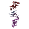
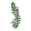
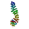
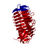
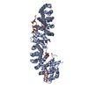
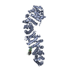
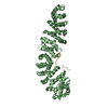
Structure of the UbcH5 :CHIP U-box complex
Components
STIP1 homology and U-Box containing protein 1
Ubiquitin-conjugating enzyme E2D 1
Keywords
LIGASE / protein-protein complex
Function / homology
Function and homology information
Oxygen-dependent proline hydroxylation of Hypoxia-inducible Factor Alpha / : / : / : / : / : / Conversion from APC/C:Cdc20 to APC/C:Cdh1 in late anaphase / Regulation of APC/C activators between G1/S and early anaphase / : / : ...Oxygen-dependent proline hydroxylation of Hypoxia-inducible Factor Alpha / : / : / : / : / : / Conversion from APC/C:Cdc20 to APC/C:Cdh1 in late anaphase / Regulation of APC/C activators between G1/S and early anaphase / : / : / Signaling by BMP / Downregulation of SMAD2/3:SMAD4 transcriptional activity / Senescence-Associated Secretory Phenotype (SASP) / CDK-mediated phosphorylation and removal of Cdc6 / Synthesis of active ubiquitin: roles of E1 and E2 enzymes / E3 ubiquitin ligases ubiquitinate target proteins / Peroxisomal protein import / Antigen processing: Ubiquitination & Proteasome degradation / : / : / : / Ovarian tumor domain proteases / Neddylation / cellular response to misfolded protein / protein quality control for misfolded or incompletely synthesized proteins / positive regulation of proteolysis / ubiquitin conjugating enzyme activity / protein K48-linked ubiquitination / ubiquitin ligase complex / positive regulation of protein ubiquitination / RING-type E3 ubiquitin transferase / Z disc / protein polyubiquitination / ubiquitin protein ligase activity / protein-folding chaperone binding / ubiquitin-dependent protein catabolic process / proteasome-mediated ubiquitin-dependent protein catabolic process / ubiquitin protein ligase binding / ATP binding / nucleus / cytoplasm Similarity search - Function
In the structure databanks used in Yorodumi, some data are registered as the other names, "COVID-19 virus" and "2019-nCoV". Here are the details of the virus and the list of structure data.
Jan 31, 2019. EMDB accession codes are about to change! (news from PDBe EMDB page)
EMDB accession codes are about to change! (news from PDBe EMDB page)
The allocation of 4 digits for EMDB accession codes will soon come to an end. Whilst these codes will remain in use, new EMDB accession codes will include an additional digit and will expand incrementally as the available range of codes is exhausted. The current 4-digit format prefixed with “EMD-” (i.e. EMD-XXXX) will advance to a 5-digit format (i.e. EMD-XXXXX), and so on. It is currently estimated that the 4-digit codes will be depleted around Spring 2019, at which point the 5-digit format will come into force.
The EM Navigator/Yorodumi systems omit the EMD- prefix.
Related info.:Q: What is EMD? / ID/Accession-code notation in Yorodumi/EM Navigator
Yorodumi is a browser for structure data from EMDB, PDB, SASBDB, etc.
This page is also the successor to EM Navigator detail page, and also detail information page/front-end page for Omokage search.
The word "yorodu" (or yorozu) is an old Japanese word meaning "ten thousand". "mi" (miru) is to see.
Related info.:EMDB / PDB / SASBDB / Comparison of 3 databanks / Yorodumi Search / Aug 31, 2016. New EM Navigator & Yorodumi / Yorodumi Papers / Jmol/JSmol / Function and homology information / Changes in new EM Navigator and Yorodumi
 Movie
Movie Controller


 Open data
 Basic information
 Components
 Keywords
 LIGASE / protein-protein complex
 Function and homology information
 Authors
 Citation
 Structure visualization
 Downloads & links
 Other downloads


 PDBj
PDBj


 Assembly
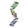


 Mass: 35.453 Da / Num. of mol.: 3 / Source method: obtained synthetically / Formula: Cl
 Mass: 18.015 Da / Num. of mol.: 4 / Source method: isolated from a natural source / Formula: H2O
 Sample preparation
 / Beamline: 4.2.2 / Wavelength: 1.24 Å
 Processing