 Movie
Movie Controller

[English] 日本語
 Yorodumi
- PDB-2ot0: Fructose-1,6-bisphosphate aldolase from rabbit muscle in complex ... -
+ Open data

- Basic information
| Entry | Database: PDB / ID: 2ot0 | ||||||
|---|---|---|---|---|---|---|---|
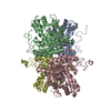
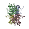
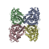
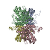
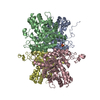
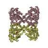
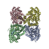
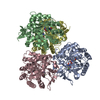
| Title | Fructose-1,6-bisphosphate aldolase from rabbit muscle in complex with a C-terminal peptide of Wiskott-Aldrich syndrome protein | ||||||
 Components Components |
| ||||||
 Keywords Keywords |  LYASE / complex / glycolysis / actin dynamics / hydrophobic pocket / WASp LYASE / complex / glycolysis / actin dynamics / hydrophobic pocket / WASp | ||||||
| Function / homology |  Function and homology information Function and homology informationregulation of T cell antigen processing and presentation / regulation of actin polymerization or depolymerization / Cdc42 protein signal transduction / GTPase regulator activity / actin filament-based movement / negative regulation of Arp2/3 complex-mediated actin nucleation / fructose-bisphosphate aldolase / fructose-bisphosphate aldolase activity / negative regulation of cell motility / vesicle membrane ...regulation of T cell antigen processing and presentation / regulation of actin polymerization or depolymerization / Cdc42 protein signal transduction / GTPase regulator activity / actin filament-based movement / negative regulation of Arp2/3 complex-mediated actin nucleation / fructose-bisphosphate aldolase / fructose-bisphosphate aldolase activity / negative regulation of cell motility / vesicle membrane / actin polymerization or depolymerization / regulation of stress fiber assembly / M band / regulation of lamellipodium assembly / I band / negative regulation of stress fiber assembly / endosomal transport / positive regulation of double-strand break repair via homologous recombination / RHOJ GTPase cycle / phospholipase binding / CDC42 GTPase cycle / Generation of second messenger molecules / RHO GTPases Activate WASPs and WAVEs / epidermis development / phagocytic vesicle / RAC1 GTPase cycle / actin filament polymerization / T cell activation / actin filament / FCGR3A-mediated phagocytosis / glycolytic process / defense response / Regulation of actin dynamics for phagocytic cup formation / small GTPase binding / SH3 domain binding / cellular response to type II interferon / cell-cell junction / blood coagulation / actin cytoskeleton / site of double-strand break / actin binding / protein homotetramerization / protein-containing complex assembly / positive regulation of cell migration / immune response / protein kinase binding / positive regulation of transcription by RNA polymerase II / extracellular exosome / identical protein binding / nucleus / plasma membrane / cytosolSimilarity search - Function | ||||||
| Biological species |  Oryctolagus cuniculus (rabbit) Oryctolagus cuniculus (rabbit) | ||||||
| Method | X-RAY DIFFRACTION / SYNCHROTRON / MOLECULAR REPLACEMENT / Resolution: 2.05 Å | ||||||
 Authors Authors | St-Jean, M. / Izard, T. / Sygusch, J. | ||||||
 Citation Citation | Journal: J.Biol.Chem. / Year: 2007 Title: A hydrophobic pocket in the active site of glycolytic aldolase mediates interactions with wiskott-Aldrich syndrome protein. Authors: St-Jean, M. / Izard, T. / Sygusch, J. | ||||||
| History |
|
- Structure visualization
| Structure viewer | Molecule: MolmilJmol/JSmol |
|---|
- Downloads & links
-Download
| PDBx/mmCIF format | 2ot0.cif.gz | 316.1 KB | Display | PDBx/mmCIF format |
|---|---|---|---|---|
| PDB format | pdb2ot0.ent.gz | 255.3 KB | Display | PDB format |
| PDBx/mmJSON format | 2ot0.json.gz | Tree view | PDBx/mmJSON format | |
| Others |  Other downloads Other downloads |
-Validation report
| Arichive directory | https://data.pdbj.org/pub/pdb/validation_reports/ot/2ot0ftp://data.pdbj.org/pub/pdb/validation_reports/ot/2ot0 | HTTPS FTP |
|---|
-Related structure data
| Related structure data | 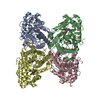 2ot1C 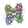 1zahS C: citing same article ( S: Starting model for refinement |
|---|---|
| Similar structure data |
-Links
 PDBj
PDBj


- Assembly
| Deposited unit | 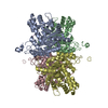
| ||||||||
|---|---|---|---|---|---|---|---|---|---|
| 1 |
| ||||||||
| Unit cell |
| ||||||||
| Details | The biological assembly is the homotetramer found in the asymmetric unit |
-Components
| #1: Protein | Mass: 39263.672 Da / Num. of mol.: 4 Source method: isolated from a genetically manipulated source Source: (gene. exp.) Oryctolagus cuniculus (rabbit) / Gene: ALDOA / Plasmid: pPB14 / Production host:  Escherichia coli (E. coli) / Strain (production host): BL21 SI / References: UniProt: P00883, fructose-bisphosphate aldolase Escherichia coli (E. coli) / Strain (production host): BL21 SI / References: UniProt: P00883, fructose-bisphosphate aldolase#2: Protein/peptide | Mass: 1782.553 Da / Num. of mol.: 4 / Source method: obtained synthetically Details: The sequence of the peptide occurs naturally in human Wiskott-Aldrich syndrome protein References: UniProt: P42768 #3: Water | ChemComp-HOH / | Water Mass: 18.015 Da / Num. of mol.: 1801 / Source method: isolated from a natural source / Formula: H2O Mass: 18.015 Da / Num. of mol.: 1801 / Source method: isolated from a natural source / Formula: H2O |
|---|
-Experimental details
-Experiment
| Experiment | Method: X-RAY DIFFRACTION / Number of used crystals: 1 |
|---|
- Sample preparation
| Crystal | Density Matthews: 2.25 Å3/Da / Density % sol: 45.23 % |
|---|---|
| Crystal grow | Temperature: 298 K / Method: vapor diffusion, hanging drop / pH: 7.5 Details: HEPES, MgCl2, PEG 550 MME, pH 7.5, VAPOR DIFFUSION, HANGING DROP, temperature 298K |
-Data collection
| Diffraction | Mean temperature: 100 K |
|---|---|
| Diffraction source | Source: SYNCHROTRON / Site: APS  / Beamline: 22-BM / Wavelength: 1 / Beamline: 22-BM / Wavelength: 1 |
| Detector | Date: Jun 7, 2006 |
| Radiation | Protocol: SINGLE WAVELENGTH / Monochromatic (M) / Laue (L): M / Scattering type: x-ray |
| Radiation wavelength | Wavelength: 1 Å / Relative weight: 1 |
| Reflection | Resolution: 2.05→50 Å / Num. all: 91889 / Num. obs: 91889 / % possible obs: 99.8 % / Redundancy: 3.4 % / Rsym value: 0.105 / Net I/σ(I): 13.1 |
| Reflection shell | Resolution: 2.05→2.16 Å / Redundancy: 3.1 % / Mean I/σ(I) obs: 2.2 / Num. unique all: 13046 / Rsym value: 0.459 / % possible all: 99.9 |
- Processing
| Software |
| |||||||||||||||||||||||||
|---|---|---|---|---|---|---|---|---|---|---|---|---|---|---|---|---|---|---|---|---|---|---|---|---|---|---|
| Refinement | Method to determine structure: MOLECULAR REPLACEMENT Starting model: PDB ENTRY 1ZAH Resolution: 2.05→50 Å / Isotropic thermal model: anisotropic / Cross valid method: THROUGHOUT / σ(I): 1
| |||||||||||||||||||||||||
| Displacement parameters |
| |||||||||||||||||||||||||
| Refine analyze |
| |||||||||||||||||||||||||
| Refinement step | Cycle: LAST / Resolution: 2.05→50 Å
| |||||||||||||||||||||||||
| Refine LS restraints |
| |||||||||||||||||||||||||
| LS refinement shell | Resolution: 2.05→2.16 Å
|
