 Movie
Movie Controller

[English] 日本語
 Yorodumi
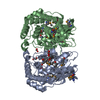
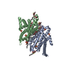
- PDB-2osu: Probable glutaminase from Bacillus subtilis complexed with 6-diaz... -
+ Open data

- Basic information
| Entry | Database: PDB / ID: 2osu | ||||||
|---|---|---|---|---|---|---|---|
| Title | Probable glutaminase from Bacillus subtilis complexed with 6-diazo-5-oxo-L-norleucine | ||||||
 Components Components | Glutaminase 1 | ||||||
 Keywords Keywords | HYDROLASE / alpha-beta structure / Structural Genomics / PSI-2 / Protein Structure Initiative / Midwest Center for Structural Genomics / MCSG | ||||||
| Function / homology |  Function and homology information Function and homology informationglutamine catabolic process / glutamate biosynthetic process / glutaminase / glutaminase activitySimilarity search - Function | ||||||
| Biological species |  Bacillus subtilis (bacteria) Bacillus subtilis (bacteria) | ||||||
| Method | X-RAY DIFFRACTION / SYNCHROTRON / SAD / Resolution: 2.29 Å | ||||||
 Authors Authors | Kim, Y. / Dementieva, I. / Vinokour, E. / Collart, F. / Joachimiak, A. / Midwest Center for Structural Genomics (MCSG) | ||||||
 Citation Citation | Journal: To be Published Title: The structure of probable glutaminase from B. subtilis complexed with its inhibitor 6-diazo-5-oxo-L-norleucine Authors: Kim, Y. / Dementieva, I. / Vinokour, E. / Collart, F. / Joachimiak, A. | ||||||
| History |
|
- Structure visualization
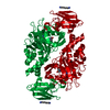
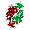
| Structure viewer | Molecule: MolmilJmol/JSmol |
|---|
- Downloads & links
-Download
| PDBx/mmCIF format | 2osu.cif.gz | 130.5 KB | Display | PDBx/mmCIF format |
|---|---|---|---|---|
| PDB format | pdb2osu.ent.gz | 109 KB | Display | PDB format |
| PDBx/mmJSON format | 2osu.json.gz | Tree view | PDBx/mmJSON format | |
| Others |  Other downloads Other downloads |
-Validation report
| Arichive directory | https://data.pdbj.org/pub/pdb/validation_reports/os/2osuftp://data.pdbj.org/pub/pdb/validation_reports/os/2osu | HTTPS FTP |
|---|
-Related structure data
| Related structure data | |
|---|---|
| Similar structure data | |
| Other databases |


-Links
 PDBj
PDBj- Assembly
| Deposited unit | 
| ||||||||
|---|---|---|---|---|---|---|---|---|---|
| 1 |
| ||||||||
| Unit cell |
|
-Components
| #1: Protein | Mass: 37059.539 Da / Num. of mol.: 2 Source method: isolated from a genetically manipulated source Source: (gene. exp.) Bacillus subtilis (bacteria) / Strain: 168 / Gene: glsA1, glsA, BSU02430 / Plasmid: pMCSG7 / Species (production host): Escherichia coli / Production host: Escherichia coli BL21(DE3) (bacteria) / Strain (production host): BL21(DE3) / References: UniProt: O31465, glutaminase#2: Chemical |   Type: L-peptide linking / Mass: 173.170 Da / Num. of mol.: 2 / Source method: obtained synthetically / Formula: C6H11N3O3 Type: L-peptide linking / Mass: 173.170 Da / Num. of mol.: 2 / Source method: obtained synthetically / Formula: C6H11N3O3#3: Water | ChemComp-HOH / | Water Mass: 18.015 Da / Num. of mol.: 232 / Source method: isolated from a natural source / Formula: H2O Mass: 18.015 Da / Num. of mol.: 232 / Source method: isolated from a natural source / Formula: H2O |
|---|
-Experimental details
-Experiment
| Experiment | Method: X-RAY DIFFRACTION / Number of used crystals: 1 |
|---|
- Sample preparation
| Crystal | Density Matthews: 2.28 Å3/Da / Density % sol: 46.08 % |
|---|---|
| Crystal grow | Temperature: 295 K / Method: vapor diffusion, hanging drop / pH: 7.5 Details: 14.5% PEG1500, 12% Glycerol, 500 mM NaCl, pH 7.5, VAPOR DIFFUSION, HANGING DROP, temperature 295K |
-Data collection
| Diffraction | Mean temperature: 100 K |
|---|---|
| Diffraction source | Source: SYNCHROTRON / Site: APS  / Beamline: 19-ID / Wavelength: 0.9793 Å / Beamline: 19-ID / Wavelength: 0.9793 Å |
| Detector | Type: SBC-2 / Detector: CCD / Date: Jul 15, 2004 / Details: mirrors |
| Radiation | Monochromator: double crystal / Protocol: SINGLE WAVELENGTH / Monochromatic (M) / Laue (L): M / Scattering type: x-ray |
| Radiation wavelength | Wavelength: 0.9793 Å / Relative weight: 1 |
| Reflection | Resolution: 2.29→34.52 Å / Num. all: 30785 / Num. obs: 30785 / % possible obs: 97.6 % / Observed criterion σ(F): 0 / Observed criterion σ(I): 0 / Redundancy: 6.3 % / Rmerge(I) obs: 0.138 / Net I/σ(I): 5.2 |
| Reflection shell | Resolution: 2.29→2.38 Å / Redundancy: 4 % / Rmerge(I) obs: 0.521 / Mean I/σ(I) obs: 1.91 / Num. unique all: 2616 / % possible all: 83.8 |
- Processing
| Software |
| ||||||||||||||||||||||||||||||||||||||||||||||||||||||||||||||||||||||||||||||||||||||||||||||||||||||||||||||||||||||||||||||||||||||||||||||||||||||||||||||||||||||||||
|---|---|---|---|---|---|---|---|---|---|---|---|---|---|---|---|---|---|---|---|---|---|---|---|---|---|---|---|---|---|---|---|---|---|---|---|---|---|---|---|---|---|---|---|---|---|---|---|---|---|---|---|---|---|---|---|---|---|---|---|---|---|---|---|---|---|---|---|---|---|---|---|---|---|---|---|---|---|---|---|---|---|---|---|---|---|---|---|---|---|---|---|---|---|---|---|---|---|---|---|---|---|---|---|---|---|---|---|---|---|---|---|---|---|---|---|---|---|---|---|---|---|---|---|---|---|---|---|---|---|---|---|---|---|---|---|---|---|---|---|---|---|---|---|---|---|---|---|---|---|---|---|---|---|---|---|---|---|---|---|---|---|---|---|---|---|---|---|---|---|---|---|
| Refinement | Method to determine structure: SAD / Resolution: 2.29→34.52 Å / Cor.coef. Fo:Fc: 0.946 / Cor.coef. Fo:Fc free: 0.91 / SU B: 14.674 / SU ML: 0.177 / Cross valid method: THROUGHOUT / σ(F): 0 / σ(I): 0 / ESU R: 0.379 / ESU R Free: 0.257 Stereochemistry target values: MAXIMUM LIKELIHOOD WITH PHASES Details: positions of DON atoms are only about 60 % confident
| ||||||||||||||||||||||||||||||||||||||||||||||||||||||||||||||||||||||||||||||||||||||||||||||||||||||||||||||||||||||||||||||||||||||||||||||||||||||||||||||||||||||||||
| Solvent computation | Ion probe radii: 0.8 Å / Shrinkage radii: 0.8 Å / VDW probe radii: 1.4 Å / Solvent model: MASK | ||||||||||||||||||||||||||||||||||||||||||||||||||||||||||||||||||||||||||||||||||||||||||||||||||||||||||||||||||||||||||||||||||||||||||||||||||||||||||||||||||||||||||
| Displacement parameters | Biso mean: 32.281 Å2
| ||||||||||||||||||||||||||||||||||||||||||||||||||||||||||||||||||||||||||||||||||||||||||||||||||||||||||||||||||||||||||||||||||||||||||||||||||||||||||||||||||||||||||
| Refinement step | Cycle: LAST / Resolution: 2.29→34.52 Å
| ||||||||||||||||||||||||||||||||||||||||||||||||||||||||||||||||||||||||||||||||||||||||||||||||||||||||||||||||||||||||||||||||||||||||||||||||||||||||||||||||||||||||||
| Refine LS restraints |
| ||||||||||||||||||||||||||||||||||||||||||||||||||||||||||||||||||||||||||||||||||||||||||||||||||||||||||||||||||||||||||||||||||||||||||||||||||||||||||||||||||||||||||
| LS refinement shell | Resolution: 2.29→2.34 Å / Total num. of bins used: 20
|
