 Movie
Movie Controller

[English] 日本語
 Yorodumi
- PDB-2oiv: Structural Analysis of Xanthomonas XopD Provides Insights Into Su... -
+ Open data

- Basic information
| Entry | Database: PDB / ID: 2oiv | ||||||
|---|---|---|---|---|---|---|---|
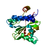
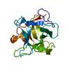
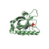
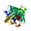
| Title | Structural Analysis of Xanthomonas XopD Provides Insights Into Substrate Specificity of Ubiquitin-like Protein Proteases | ||||||
 Components Components | Xanthomonas outer protein D | ||||||
 Keywords Keywords |  HYDROLASE / Clan CE Family 48 Cysteine protease / Type III secreted effector / deSUMOylating enzyme / secreted virulence factor / peptidase / isopeptidase HYDROLASE / Clan CE Family 48 Cysteine protease / Type III secreted effector / deSUMOylating enzyme / secreted virulence factor / peptidase / isopeptidase | ||||||
| Function / homology |  Function and homology information Function and homology information | ||||||
| Biological species |  Xanthomonas euvesicatoria (bacteria) Xanthomonas euvesicatoria (bacteria) | ||||||
| Method | X-RAY DIFFRACTION / SYNCHROTRON / SAD / Resolution: 1.95 Å | ||||||
 Authors Authors | Chosed, R. / Tomchick, D.R. / Brautigam, C.A. / Machius, M. / Orth, K. | ||||||
 Citation Citation | Journal: J.Biol.Chem. / Year: 2007 Title: Structural analysis of Xanthomonas XopD provides insights into substrate specificity of ubiquitin-like protein proteases. Authors: Chosed, R. / Tomchick, D.R. / Brautigam, C.A. / Mukherjee, S. / Negi, V.S. / Machius, M. / Orth, K. #1: Journal: Biochem.J. / Year: 2006Title: Evolution of a signalling system that incorporates both redundancy and diversity: Arabidopsis SUMOylation. Authors: Chosed, R. / Mukherjee, S. / Lois, L.M. / Orth, K. #2: Journal: Mol.Microbiol. / Year: 2003Title: Xanthomonas type III effector XopD targets SUMO-conjugated proteins in planta. Authors: Hotson, A. / Chosed, R. / Shu, H. / Orth, K. / Mudgett, M.B. #3: Journal: Mol.Cell / Year: 2000Title: Ulp1-SUMO Crystal Structure and Genetic Analysis Reveal Conserved Interactions and a Regulatory Element Essential for Cell Growth in Yeast Authors: Mossessova, E. / Lima, C.D. | ||||||
| History |
|
- Structure visualization
| Structure viewer | Molecule: MolmilJmol/JSmol |
|---|
- Downloads & links
-Download
| PDBx/mmCIF format | 2oiv.cif.gz | 49.8 KB | Display | PDBx/mmCIF format |
|---|---|---|---|---|
| PDB format | pdb2oiv.ent.gz | 35.7 KB | Display | PDB format |
| PDBx/mmJSON format | 2oiv.json.gz | Tree view | PDBx/mmJSON format | |
| Others |  Other downloads Other downloads |
-Validation report
| Arichive directory | https://data.pdbj.org/pub/pdb/validation_reports/oi/2oivftp://data.pdbj.org/pub/pdb/validation_reports/oi/2oiv | HTTPS FTP |
|---|
-Related structure data


-Links
 PDBj
PDBj- Assembly
| Deposited unit | 
| ||||||||
|---|---|---|---|---|---|---|---|---|---|
| 1 |
| ||||||||
| Unit cell |
|
-Components
| #1: Protein | Mass: 20763.115 Da / Num. of mol.: 1 / Fragment: catalytic fragment (Residues 335-520) Source method: isolated from a genetically manipulated source Source: (gene. exp.) Xanthomonas euvesicatoria (bacteria) / Gene: xopD / Plasmid: pGEX-rTEV / Species (production host): Escherichia coli / Production host: Escherichia coli BL21(DE3) (bacteria) / Strain (production host): BL21/DE3 / References: UniProt: Q3BYJ5 |
|---|---|
| #2: Chemical | ChemComp-PO4 / Phosphate  Mass: 94.971 Da / Num. of mol.: 1 / Source method: obtained synthetically / Formula: PO4 Mass: 94.971 Da / Num. of mol.: 1 / Source method: obtained synthetically / Formula: PO4 |
| #3: Water | ChemComp-HOH / Water Mass: 18.015 Da / Num. of mol.: 83 / Source method: isolated from a natural source / Formula: H2O Mass: 18.015 Da / Num. of mol.: 83 / Source method: isolated from a natural source / Formula: H2O |
-Experimental details
-Experiment
| Experiment | Method: X-RAY DIFFRACTION / Number of used crystals: 1 |
|---|
- Sample preparation
| Crystal | Density Matthews: 2.26 Å3/Da / Density % sol: 45.67 % |
|---|---|
| Crystal grow | Temperature: 298 K / Method: vapor diffusion, hanging drop / pH: 7.5 Details: 15 mg/mL protein in 20 mM Tris-HCl pH 7.5, 75 mM KCl, and 0.5 mM DTT, 1.4 - 1.6 M sodium potassium phosphate, VAPOR DIFFUSION, HANGING DROP, temperature 298.0K |
-Data collection
| Diffraction | Mean temperature: 100 K |
|---|---|
| Diffraction source | Source: SYNCHROTRON / Site: APS  / Beamline: 19-BM / Wavelength: 0.97929 Å / Beamline: 19-BM / Wavelength: 0.97929 Å |
| Detector | Type: SBC-3 / Detector: CCD / Date: Mar 17, 2004 |
| Radiation | Monochromator: NONE / Protocol: SINGLE WAVELENGTH / Monochromatic (M) / Laue (L): M / Scattering type: x-ray |
| Radiation wavelength | Wavelength: 0.97929 Å / Relative weight: 1 |
| Reflection | Resolution: 1.95→32.04 Å / Num. all: 14282 / Num. obs: 14282 / % possible obs: 98.6 % / Observed criterion σ(F): 0 / Observed criterion σ(I): 0 / Redundancy: 7.6 % / Biso Wilson estimate: 37.8 Å2 / Rmerge(I) obs: 0.061 / Net I/σ(I): 31.2 |
| Reflection shell | Resolution: 1.95→2 Å / Redundancy: 7.5 % / Rmerge(I) obs: 0.71 / Mean I/σ(I) obs: 2.25 / % possible all: 99.9 |
- Processing
| Software |
| |||||||||||||||||||||||||||||||||||||||||||||||||||||||||||||||||||||||||||||||||||||||||||||||||||||||||||||||||||||||||||||||||||||||||||||||||||||||||||||||||||||||||||||||
|---|---|---|---|---|---|---|---|---|---|---|---|---|---|---|---|---|---|---|---|---|---|---|---|---|---|---|---|---|---|---|---|---|---|---|---|---|---|---|---|---|---|---|---|---|---|---|---|---|---|---|---|---|---|---|---|---|---|---|---|---|---|---|---|---|---|---|---|---|---|---|---|---|---|---|---|---|---|---|---|---|---|---|---|---|---|---|---|---|---|---|---|---|---|---|---|---|---|---|---|---|---|---|---|---|---|---|---|---|---|---|---|---|---|---|---|---|---|---|---|---|---|---|---|---|---|---|---|---|---|---|---|---|---|---|---|---|---|---|---|---|---|---|---|---|---|---|---|---|---|---|---|---|---|---|---|---|---|---|---|---|---|---|---|---|---|---|---|---|---|---|---|---|---|---|---|---|
| Refinement | Method to determine structure: SAD / Resolution: 1.95→32.04 Å / Cor.coef. Fo:Fc: 0.952 / Cor.coef. Fo:Fc free: 0.919 / SU B: 9.92 / SU ML: 0.141 / TLS residual ADP flag: LIKELY RESIDUAL / Isotropic thermal model: isotropic / Cross valid method: THROUGHOUT / σ(F): 0 / σ(I): 0 / ESU R: 0.177 / ESU R Free: 0.162 / Stereochemistry target values: MAXIMUM LIKELIHOOD / Details: HYDROGENS HAVE BEEN ADDED IN THE RIDING POSITIONS
| |||||||||||||||||||||||||||||||||||||||||||||||||||||||||||||||||||||||||||||||||||||||||||||||||||||||||||||||||||||||||||||||||||||||||||||||||||||||||||||||||||||||||||||||
| Solvent computation | Ion probe radii: 0.8 Å / Shrinkage radii: 0.8 Å / VDW probe radii: 1.4 Å / Solvent model: BABINET MODEL WITH MASK | |||||||||||||||||||||||||||||||||||||||||||||||||||||||||||||||||||||||||||||||||||||||||||||||||||||||||||||||||||||||||||||||||||||||||||||||||||||||||||||||||||||||||||||||
| Displacement parameters | Biso mean: 42.341 Å2
| |||||||||||||||||||||||||||||||||||||||||||||||||||||||||||||||||||||||||||||||||||||||||||||||||||||||||||||||||||||||||||||||||||||||||||||||||||||||||||||||||||||||||||||||
| Refine analyze |
| |||||||||||||||||||||||||||||||||||||||||||||||||||||||||||||||||||||||||||||||||||||||||||||||||||||||||||||||||||||||||||||||||||||||||||||||||||||||||||||||||||||||||||||||
| Refinement step | Cycle: LAST / Resolution: 1.95→32.04 Å
| |||||||||||||||||||||||||||||||||||||||||||||||||||||||||||||||||||||||||||||||||||||||||||||||||||||||||||||||||||||||||||||||||||||||||||||||||||||||||||||||||||||||||||||||
| Refine LS restraints |
| |||||||||||||||||||||||||||||||||||||||||||||||||||||||||||||||||||||||||||||||||||||||||||||||||||||||||||||||||||||||||||||||||||||||||||||||||||||||||||||||||||||||||||||||
| LS refinement shell | Resolution: 1.95→2 Å / Total num. of bins used: 20
| |||||||||||||||||||||||||||||||||||||||||||||||||||||||||||||||||||||||||||||||||||||||||||||||||||||||||||||||||||||||||||||||||||||||||||||||||||||||||||||||||||||||||||||||
| Refinement TLS params. | Method: refined / Refine-ID: X-RAY DIFFRACTION
| |||||||||||||||||||||||||||||||||||||||||||||||||||||||||||||||||||||||||||||||||||||||||||||||||||||||||||||||||||||||||||||||||||||||||||||||||||||||||||||||||||||||||||||||
| Refinement TLS group |
|
