 Movie
Movie Controller

[English] 日本語
 Yorodumi
- PDB-2ohe: Structural and mutational analysis of tRNA-Intron splicing endonu... -
+ Open data

- Basic information
| Entry | Database: PDB / ID: 2ohe | ||||||
|---|---|---|---|---|---|---|---|
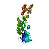
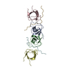
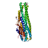
| Title | Structural and mutational analysis of tRNA-Intron splicing endonuclease from Thermoplasma acidophilum DSM 1728 | ||||||
 Components Components | tRNA-splicing endonuclease | ||||||
 Keywords Keywords |  HYDROLASE / tRNA / Intron / splicing / endonuclease HYDROLASE / tRNA / Intron / splicing / endonuclease | ||||||
| Function / homology |  Function and homology information Function and homology informationtRNA-type intron splice site recognition and cleavage / tRNA-intron lyase / tRNA-intron endonuclease activity / nucleic acid binding / lyase activitySimilarity search - Function | ||||||
| Biological species |   Thermoplasma acidophilum (acidophilic) Thermoplasma acidophilum (acidophilic) | ||||||
| Method | X-RAY DIFFRACTION / SYNCHROTRON / MAD / Resolution: 2.7 Å | ||||||
 Authors Authors | Kim, Y.K. / Mizutani, K. / Rhee, K.H. / Lee, W.H. / Park, S.Y. / Hwang, K.Y. | ||||||
 Citation Citation | Journal: J.Bacteriol. / Year: 2007 Title: Structural and Mutational Analysis of tRNA Intron-Splicing Endonuclease from Thermoplasma acidophilum DSM 1728: Catalytic Mechanism of tRNA Intron-Splicing Endonucleases Authors: Kim, Y.K. / Mizutani, K. / Rhee, K.H. / Nam, K.H. / Lee, W.H. / Lee, E.H. / Kim, E.E. / Park, S.Y. / Hwang, K.Y. | ||||||
| History |
|
- Structure visualization
| Structure viewer | Molecule: MolmilJmol/JSmol |
|---|
- Downloads & links
-Download
| PDBx/mmCIF format | 2ohe.cif.gz | 73.1 KB | Display | PDBx/mmCIF format |
|---|---|---|---|---|
| PDB format | pdb2ohe.ent.gz | 54.2 KB | Display | PDB format |
| PDBx/mmJSON format | 2ohe.json.gz | Tree view | PDBx/mmJSON format | |
| Others |  Other downloads Other downloads |
-Validation report
| Arichive directory | https://data.pdbj.org/pub/pdb/validation_reports/oh/2oheftp://data.pdbj.org/pub/pdb/validation_reports/oh/2ohe | HTTPS FTP |
|---|
-Related structure data


-Links
 PDBj
PDBj- Assembly
| Deposited unit | 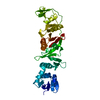
| ||||||||
|---|---|---|---|---|---|---|---|---|---|
| 1 |
| ||||||||
| 2 | 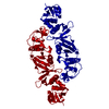
| ||||||||
| Unit cell |
|
-Components
| #1: Protein | Mass: 33394.777 Da / Num. of mol.: 1 Source method: isolated from a genetically manipulated source Source: (gene. exp.) Thermoplasma acidophilum (acidophilic) / Strain: DSM 1728 / Gene: endA / Plasmid: pET28a / Species (production host): Escherichia coli / Production host:  Escherichia coli BL21(DE3) (bacteria) / Strain (production host): BL 21 DE3 / References: UniProt: Q9HIY5, EC: 3.1.27.9 Escherichia coli BL21(DE3) (bacteria) / Strain (production host): BL 21 DE3 / References: UniProt: Q9HIY5, EC: 3.1.27.9 |
|---|---|
| #2: Water | ChemComp-HOH / Water Mass: 18.015 Da / Num. of mol.: 116 / Source method: isolated from a natural source / Formula: H2O Mass: 18.015 Da / Num. of mol.: 116 / Source method: isolated from a natural source / Formula: H2O |
-Experimental details
-Experiment
| Experiment | Method: X-RAY DIFFRACTION / Number of used crystals: 1 |
|---|
- Sample preparation
| Crystal | Density Matthews: 3.3 Å3/Da / Density % sol: 62.68 % |
|---|---|
| Crystal grow | Temperature: 295 K / Method: vapor diffusion, hanging drop / pH: 6.2 Details: 2.4M NaCl, 0.1M Na/K phosphate, pH 6.2, VAPOR DIFFUSION, HANGING DROP, temperature 295K |
-Data collection
| Diffraction | Mean temperature: 100 K |
|---|---|
| Diffraction source | Source: SYNCHROTRON / Site: PAL/PLS  / Beamline: 4A / Wavelength: 0.9795 Å / Beamline: 4A / Wavelength: 0.9795 Å |
| Detector | Type: ADSC QUANTUM 210 / Detector: CCD / Date: Feb 12, 2006 / Details: wiggler |
| Radiation | Monochromator: wiggler / Protocol: MAD / Monochromatic (M) / Laue (L): M / Scattering type: x-ray |
| Radiation wavelength | Wavelength: 0.9795 Å / Relative weight: 1 |
| Reflection | Resolution: 2.7→50 Å / Num. obs: 35520 / % possible obs: 98 % / Observed criterion σ(F): 2 / Observed criterion σ(I): 0 / Biso Wilson estimate: 32.6 Å2 / Rsym value: 0.066 |
| Reflection shell | Resolution: 2.7→2.9 Å / Mean I/σ(I) obs: 4.44 / Num. unique all: 1887 / Rsym value: 0.29 / % possible all: 97.6 |
- Processing
| Software |
| |||||||||||||||||||||||||
|---|---|---|---|---|---|---|---|---|---|---|---|---|---|---|---|---|---|---|---|---|---|---|---|---|---|---|
| Refinement | Method to determine structure: MAD / Resolution: 2.7→17.79 Å / Rfactor Rfree error: 0.009 / Data cutoff high absF: 273973.83 / Data cutoff low absF: 0 / Isotropic thermal model: RESTRAINED / Cross valid method: THROUGHOUT / σ(F): 2 / Stereochemistry target values: Engh & Huber
| |||||||||||||||||||||||||
| Solvent computation | Solvent model: FLAT MODEL / Bsol: 46.3258 Å2 / ksol: 0.385537 e/Å3 | |||||||||||||||||||||||||
| Displacement parameters | Biso mean: 44.5 Å2
| |||||||||||||||||||||||||
| Refine analyze |
| |||||||||||||||||||||||||
| Refinement step | Cycle: LAST / Resolution: 2.7→17.79 Å
| |||||||||||||||||||||||||
| Refine LS restraints |
| |||||||||||||||||||||||||
| LS refinement shell | Resolution: 2.7→2.87 Å / Rfactor Rfree error: 0.04 / Total num. of bins used: 6
| |||||||||||||||||||||||||
| Xplor file |
|
