 Movie
Movie Controller

[English] 日本語
 Yorodumi
- PDB-2odl: Crystal structure of the HMW1 secretion domain from Haemophilus i... -
+ Open data

- Basic information
| Entry | Database: PDB / ID: 2odl | ||||||
|---|---|---|---|---|---|---|---|
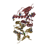
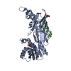
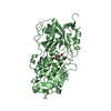
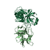
| Title | Crystal structure of the HMW1 secretion domain from Haemophilus influenzae | ||||||
 Components Components | Adhesin | ||||||
 Keywords Keywords |  CELL ADHESION / HMW1 / secretion domain / beta helix CELL ADHESION / HMW1 / secretion domain / beta helix | ||||||
| Function / homology |  Function and homology information Function and homology informationFilamentous haemagglutinin FhaB/tRNA nuclease CdiA-like, TPS domain / TPS secretion domain / haemagglutination activity domain / ESPR domain / Extended Signal Peptide of Type V secretion system / Single-stranded right-handed beta-helix, Pectin lyase-like / Pectate Lyase C-like / Pectin lyase fold / Pectin lyase fold/virulence factor / 3 Solenoid / Mainly BetaSimilarity search - Domain/homology | ||||||
| Biological species |  Haemophilus influenzae (bacteria) Haemophilus influenzae (bacteria) | ||||||
| Method | X-RAY DIFFRACTION / SYNCHROTRON / SAD / Resolution: 1.92 Å | ||||||
 Authors Authors | Yokoyama, T. / Yeo, H.J. | ||||||
 Citation Citation | Journal: J.Biol.Chem. / Year: 2007 Title: The structure of the Haemophilus influenzae HMW1 pro-piece reveals a structural domain essential for bacterial two-partner secretion. Authors: Yeo, H.J. / Yokoyama, T. / Walkiewicz, K. / Kim, Y. / Grass, S. / Geme, J.W. | ||||||
| History |
|
- Structure visualization
| Structure viewer | Molecule: MolmilJmol/JSmol |
|---|
- Downloads & links
-Download
| PDBx/mmCIF format | 2odl.cif.gz | 85.6 KB | Display | PDBx/mmCIF format |
|---|---|---|---|---|
| PDB format | pdb2odl.ent.gz | 63.3 KB | Display | PDB format |
| PDBx/mmJSON format | 2odl.json.gz | Tree view | PDBx/mmJSON format | |
| Others |  Other downloads Other downloads |
-Validation report
| Arichive directory | https://data.pdbj.org/pub/pdb/validation_reports/od/2odlftp://data.pdbj.org/pub/pdb/validation_reports/od/2odl | HTTPS FTP |
|---|
-Related structure data
| Similar structure data |
|---|


-Links
 PDBj
PDBj- Assembly
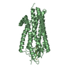
| Deposited unit | 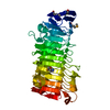
| ||||||||
|---|---|---|---|---|---|---|---|---|---|
| 1 |
| ||||||||
| Unit cell |
| ||||||||
| Details | The biological assembly is a monomer in the asymmetric unit |
-Components
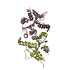
| #1: Protein | Mass: 39362.391 Da / Num. of mol.: 1 / Fragment: Residues 69-441 Source method: isolated from a genetically manipulated source Source: (gene. exp.) Haemophilus influenzae (bacteria) / Strain: 12 / Gene: hmw1A / Plasmid: pGEX6P1 / Species (production host): Escherichia coli / Production host: Escherichia coli BL21(DE3) (bacteria) / Strain (production host): BL21(DE3) / References: UniProt: Q48031 |
|---|---|
| #2: Water | ChemComp-HOH / Water Mass: 18.015 Da / Num. of mol.: 182 / Source method: isolated from a natural source / Formula: H2O Mass: 18.015 Da / Num. of mol.: 182 / Source method: isolated from a natural source / Formula: H2O |
-Experimental details
-Experiment
| Experiment | Method: X-RAY DIFFRACTION / Number of used crystals: 1 |
|---|
- Sample preparation
| Crystal | Density Matthews: 2.37 Å3/Da / Density % sol: 48.07 % |
|---|---|
| Crystal grow | Temperature: 290 K / Method: vapor diffusion, hanging drop / pH: 7.2 Details: 24% PEG6000, 0.1 M Tris-HCl, pH 7.2, VAPOR DIFFUSION, HANGING DROP, temperature 290K |
-Data collection
| Diffraction | Mean temperature: 100 K |
|---|---|
| Diffraction source | Source: SYNCHROTRON / Site: APS  / Beamline: 19-BM / Wavelength: 0.97904 Å / Beamline: 19-BM / Wavelength: 0.97904 Å |
| Detector | Type: SBC-3 / Detector: CCD / Date: Jul 5, 2006 |
| Radiation | Monochromator: Si 111 / Protocol: SINGLE WAVELENGTH / Monochromatic (M) / Laue (L): M / Scattering type: x-ray |
| Radiation wavelength | Wavelength: 0.97904 Å / Relative weight: 1 |
| Reflection | Resolution: 1.92→50 Å / Num. obs: 28035 / % possible obs: 98.8 % / Observed criterion σ(I): 0 / Redundancy: 6.5 % / Biso Wilson estimate: 17.5 Å2 / Rmerge(I) obs: 0.099 / Net I/σ(I): 6.2 |
| Reflection shell | Resolution: 1.92→1.99 Å / Redundancy: 4.3 % / Rmerge(I) obs: 0.228 / Num. unique all: 2690 / % possible all: 95.1 |
- Processing
| Software |
| ||||||||||||||||||||||||||||||||||||||||||||||||||||||||||||||||||||||||||||||||||||||||||||||||||||||||||||||||||||||||||||||||||||||||||||||||||||||||||||||||||||||||||
|---|---|---|---|---|---|---|---|---|---|---|---|---|---|---|---|---|---|---|---|---|---|---|---|---|---|---|---|---|---|---|---|---|---|---|---|---|---|---|---|---|---|---|---|---|---|---|---|---|---|---|---|---|---|---|---|---|---|---|---|---|---|---|---|---|---|---|---|---|---|---|---|---|---|---|---|---|---|---|---|---|---|---|---|---|---|---|---|---|---|---|---|---|---|---|---|---|---|---|---|---|---|---|---|---|---|---|---|---|---|---|---|---|---|---|---|---|---|---|---|---|---|---|---|---|---|---|---|---|---|---|---|---|---|---|---|---|---|---|---|---|---|---|---|---|---|---|---|---|---|---|---|---|---|---|---|---|---|---|---|---|---|---|---|---|---|---|---|---|---|---|---|
| Refinement | Method to determine structure: SAD / Resolution: 1.92→31.61 Å / Cor.coef. Fo:Fc: 0.949 / Cor.coef. Fo:Fc free: 0.922 / SU B: 2.939 / SU ML: 0.089 / Isotropic thermal model: Isotropic / Cross valid method: THROUGHOUT / σ(F): 0 / σ(I): 0 / ESU R: 0.151 / ESU R Free: 0.141 / Stereochemistry target values: MAXIMUM LIKELIHOOD
| ||||||||||||||||||||||||||||||||||||||||||||||||||||||||||||||||||||||||||||||||||||||||||||||||||||||||||||||||||||||||||||||||||||||||||||||||||||||||||||||||||||||||||
| Solvent computation | Ion probe radii: 0.8 Å / Shrinkage radii: 0.8 Å / VDW probe radii: 1.4 Å / Solvent model: MASK | ||||||||||||||||||||||||||||||||||||||||||||||||||||||||||||||||||||||||||||||||||||||||||||||||||||||||||||||||||||||||||||||||||||||||||||||||||||||||||||||||||||||||||
| Displacement parameters | Biso mean: 14.904 Å2
| ||||||||||||||||||||||||||||||||||||||||||||||||||||||||||||||||||||||||||||||||||||||||||||||||||||||||||||||||||||||||||||||||||||||||||||||||||||||||||||||||||||||||||
| Refinement step | Cycle: LAST / Resolution: 1.92→31.61 Å
| ||||||||||||||||||||||||||||||||||||||||||||||||||||||||||||||||||||||||||||||||||||||||||||||||||||||||||||||||||||||||||||||||||||||||||||||||||||||||||||||||||||||||||
| Refine LS restraints |
| ||||||||||||||||||||||||||||||||||||||||||||||||||||||||||||||||||||||||||||||||||||||||||||||||||||||||||||||||||||||||||||||||||||||||||||||||||||||||||||||||||||||||||
| LS refinement shell | Resolution: 1.92→1.97 Å / Total num. of bins used: 20
|
