[English] 日本語
Yorodumi
- PDB-2o8d: human MutSalpha (MSH2/MSH6) bound to ADP and a G dU mispair -

+
Open data


ID or keywords:

Loading...

-
Basic information

Entry
Database: PDB / ID: 2o8d
Titlehuman MutSalpha (MSH2/MSH6) bound to ADP and a G dU mispair
Components
  • (DNA mismatch repair protein ...) x 2
  • 5'-D(*CP*CP*TP*AP*GP*CP*GP*(DU)P*GP*CP*GP*GP*TP*TP*C)-3'
  • 5'-D(*GP*AP*AP*CP*CP*GP*CP*GP*CP*GP*CP*TP*AP*GP*G)-3'
Keywords
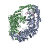
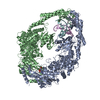
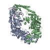
DNA BINDING PROTEIN/DNA / DNA mismatch repair / DNA damage response / somatic hypermutation / protein-DNA complex / DNA mispair / cancer / DNA BINDING PROTEIN-DNA COMPLEX
Function / homology
Function and homology information


somatic recombination of immunoglobulin genes involved in immune response / MutSbeta complex / Defective Mismatch Repair Associated With MSH3 / MutSalpha complex / Defective Mismatch Repair Associated With MSH2 / Defective Mismatch Repair Associated With MSH6 / guanine/thymine mispair binding / somatic recombination of immunoglobulin gene segments / maintenance of DNA repeat elements / B cell mediated immunity ...somatic recombination of immunoglobulin genes involved in immune response / MutSbeta complex / Defective Mismatch Repair Associated With MSH3 / MutSalpha complex / Defective Mismatch Repair Associated With MSH2 / Defective Mismatch Repair Associated With MSH6 / guanine/thymine mispair binding / somatic recombination of immunoglobulin gene segments / maintenance of DNA repeat elements / B cell mediated immunity / positive regulation of helicase activity / positive regulation of isotype switching to IgA isotypes / meiotic mismatch repair / centromeric DNA binding / positive regulation of isotype switching to IgG isotypes / mismatched DNA binding / negative regulation of DNA recombination / mitotic recombination / isotype switching / Mismatch repair (MMR) directed by MSH2:MSH3 (MutSbeta) / Mismatch repair (MMR) directed by MSH2:MSH6 (MutSalpha) / oxidative phosphorylation / postreplication repair / response to UV-B / mitotic intra-S DNA damage checkpoint signaling / ATP-dependent DNA damage sensor activity / germ cell development / response to X-ray / intrinsic apoptotic signaling pathway in response to DNA damage by p53 class mediator / somatic hypermutation of immunoglobulin genes / ATP-dependent activity, acting on DNA / mismatch repair / response to UV / protein localization to chromatin / methylated histone binding / intrinsic apoptotic signaling pathway / B cell differentiation / determination of adult lifespan / TP53 Regulates Transcription of DNA Repair Genes / male gonad development / intrinsic apoptotic signaling pathway in response to DNA damage / double-strand break repair / spermatogenesis / in utero embryonic development / negative regulation of neuron apoptotic process / damaged DNA binding / chromosome, telomeric region / intracellular membrane-bounded organelle / DNA repair / chromatin binding / chromatin / Golgi apparatus / enzyme binding / protein homodimerization activity / ATP hydrolysis activity / DNA binding / nucleoplasm / ATP binding / membrane / nucleus / cytosol
Similarity search - Function
DNA mismatch repair Msh2-type / DNA mismatch repair protein Msh2 / MutS, connector domain / DNA repair protein MutS, domain I / MutS, DNA mismatch repair protein; Chain A, domain 3 / MutS, DNA mismatch repair protein; Chain A, domain 3 - #10 / DNA mismatch repair protein MutS/MSH / DNA mismatch repair protein MutS-like, N-terminal / DNA mismatch repair protein MutS, connector domain / DNA mismatch repair protein MutS, clamp ...DNA mismatch repair Msh2-type / DNA mismatch repair protein Msh2 / MutS, connector domain / DNA repair protein MutS, domain I / MutS, DNA mismatch repair protein; Chain A, domain 3 / MutS, DNA mismatch repair protein; Chain A, domain 3 - #10 / DNA mismatch repair protein MutS/MSH / DNA mismatch repair protein MutS-like, N-terminal / DNA mismatch repair protein MutS, connector domain / DNA mismatch repair protein MutS, clamp / DNA mismatch repair protein MutS, N-terminal / MutS, connector domain superfamily / MutS domain I / MutS domain II / MutS family domain IV / MutS domain III / DNA mismatch repair MutS family / DNA mismatch repair protein MutS, C-terminal / DNA mismatch repair protein MutS, core / DNA mismatch repair protein MutS, core domain superfamily / MutS domain V / DNA mismatch repair proteins mutS family signature. / DNA-binding domain of DNA mismatch repair MUTS family / ATPase domain of DNA mismatch repair MUTS family / MutS, DNA mismatch repair protein, domain I / domain with conserved PWWP motif / PWWP domain / PWWP domain profile. / PWWP domain / Nucleotidyltransferase; domain 5 / P-loop containing nucleotide triphosphate hydrolases / P-loop containing nucleoside triphosphate hydrolase / Rossmann fold / 2-Layer Sandwich / Orthogonal Bundle / 3-Layer(aba) Sandwich / Mainly Alpha / Alpha Beta
Similarity search - Domain/homology
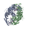
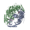
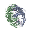
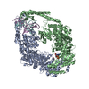
ADENOSINE-5'-DIPHOSPHATE / DNA / DNA (> 10) / DNA mismatch repair protein Msh2 / DNA mismatch repair protein Msh6
Similarity search - Component
Biological speciesHomo sapiens (human)
MethodX-RAY DIFFRACTION / SYNCHROTRON / FOURIER SYNTHESIS / Resolution: 3 Å
AuthorsWarren, J.J. / Pohlhaus, T.J. / Changela, A. / Modrich, P.L. / Beese, L.S.
CitationJournal: Mol.Cell / Year: 2007
Title: Structure of the Human MutSalpha DNA Lesion Recognition Complex.
Authors: Warren, J.J. / Pohlhaus, T.J. / Changela, A. / Iyer, R.R. / Modrich, P.L. / Beese, L.S.
History
DepositionDec 12, 2006Deposition site: RCSB / Processing site: RCSB
Revision 1.0Jun 5, 2007Provider: repository / Type: Initial release
Revision 1.1May 1, 2008Group: Version format compliance
Revision 1.2Jul 13, 2011Group: Advisory / Version format compliance
Revision 1.3Dec 27, 2023Group: Data collection / Database references / Derived calculations
Category: chem_comp_atom / chem_comp_bond ...chem_comp_atom / chem_comp_bond / database_2 / pdbx_struct_special_symmetry / struct_conn / struct_ref_seq_dif / struct_site
Item: _database_2.pdbx_DOI / _database_2.pdbx_database_accession ..._database_2.pdbx_DOI / _database_2.pdbx_database_accession / _struct_conn.ptnr1_auth_comp_id / _struct_conn.ptnr1_auth_seq_id / _struct_conn.ptnr1_label_asym_id / _struct_conn.ptnr1_label_atom_id / _struct_conn.ptnr1_label_comp_id / _struct_conn.ptnr1_label_seq_id / _struct_conn.ptnr2_auth_comp_id / _struct_conn.ptnr2_auth_seq_id / _struct_conn.ptnr2_label_asym_id / _struct_conn.ptnr2_label_atom_id / _struct_conn.ptnr2_label_comp_id / _struct_conn.ptnr2_label_seq_id / _struct_ref_seq_dif.details / _struct_site.pdbx_auth_asym_id / _struct_site.pdbx_auth_comp_id / _struct_site.pdbx_auth_seq_id

-
Structure visualization

Structure viewerMolecule:
MolmilJmol/JSmol

Downloads & links

-
Assembly

Deposited unit
E: 5'-D(*GP*AP*AP*CP*CP*GP*CP*GP*CP*GP*CP*TP*AP*GP*G)-3'
F: 5'-D(*CP*CP*TP*AP*GP*CP*GP*(DU)P*GP*CP*GP*GP*TP*TP*C)-3'
A: DNA mismatch repair protein Msh2
B: DNA mismatch repair protein MSH6
hetero molecules


Theoretical massNumber of molelcules
Total (without water)231,0048
Polymers230,1014
Non-polymers9034
Water82946
1


  • Idetical with deposited unit
  • defined by author
TypeNameSymmetry operationNumber
identity operation1_555x,y,z1
Unit cell
Length a, b, c (Å)260.379, 260.379, 260.379
Angle α, β, γ (deg.)90.00, 90.00, 90.00
Int Tables number212
Space group name H-MP4332
Components on special symmetry positions
IDModelComponents
11B-26-

HOH

-
Components

-
DNA chain , 2 types, 2 molecules EF

#1: DNA chain 5'-D(*GP*AP*AP*CP*CP*GP*CP*GP*CP*GP*CP*TP*AP*GP*G)-3'


Mass: 4620.000 Da / Num. of mol.: 1 / Source method: obtained synthetically
#2: DNA chain 5'-D(*CP*CP*TP*AP*GP*CP*GP*(DU)P*GP*CP*GP*GP*TP*TP*C)-3'


Mass: 4562.932 Da / Num. of mol.: 1 / Source method: obtained synthetically

-
DNA mismatch repair protein ... , 2 types, 2 molecules AB

#3: Protein DNA mismatch repair protein Msh2 / / MutS protein homolog 2


Mass: 104861.875 Da / Num. of mol.: 1
Source method: isolated from a genetically manipulated source
Source: (gene. exp.) Homo sapiens (human) / Gene: MSH2 / Plasmid: pFasBacDual / Production host: Spodoptera frugiperda (fall armyworm) / Strain (production host): sf9 / References: UniProt: P43246
#4: Protein DNA mismatch repair protein MSH6 / / MutS-alpha 160 kDa subunit / G/T mismatch-binding protein / GTBP / GTMBP / p160


Mass: 116056.672 Da / Num. of mol.: 1 / Fragment: residues 341-1360
Source method: isolated from a genetically manipulated source
Source: (gene. exp.) Homo sapiens (human) / Gene: MSH6, GTBP / Plasmid: pFasBacDual / Production host: Spodoptera frugiperda (fall armyworm) / Strain (production host): sf9 / References: UniProt: P52701

-
Non-polymers , 3 types, 50 molecules

#5: Chemical ChemComp-MG / MAGNESIUM ION


Mass: 24.305 Da / Num. of mol.: 2 / Source method: obtained synthetically / Formula: Mg
#6: Chemical ChemComp-ADP / ADENOSINE-5'-DIPHOSPHATE / Adenosine diphosphate


Mass: 427.201 Da / Num. of mol.: 2 / Source method: obtained synthetically / Formula: C10H15N5O10P2 / Comment: ADP, energy-carrying molecule*YM
#7: Water ChemComp-HOH / water / Water


Mass: 18.015 Da / Num. of mol.: 46 / Source method: isolated from a natural source / Formula: H2O

-
Experimental details

-
Experiment

ExperimentMethod: X-RAY DIFFRACTION / Number of used crystals: 1

-
Sample preparation

CrystalDensity Matthews: 3.19 Å3/Da / Density % sol: 61.44 %
Crystal growTemperature: 290 K / Method: vapor diffusion / pH: 7
Details: 11% PEG 8000, 10 mM magnesium sulfate, 100 mM bis-tris-propane, pH 7, VAPOR DIFFUSION, temperature 290K
Components of the solutions
IDNameCrystal-IDSol-ID
1PEG 800011
2magnesium sulfate11
3bis-tris-propane11
4HOH11
5PEG 800012
6magnesium sulfate12
7bis-tris-propane12
8HOH12

-
Data collection

DiffractionMean temperature: 100 K
Diffraction sourceSource: SYNCHROTRON / Site: APS / Beamline: 22-ID / Wavelength: 0.9793 Å
DetectorType: MARMOSAIC 300 mm CCD / Detector: CCD / Details: mirrors
RadiationMonochromator: Si 220 / Protocol: SINGLE WAVELENGTH / Monochromatic (M) / Laue (L): M / Scattering type: x-ray
Radiation wavelengthWavelength: 0.9793 Å / Relative weight: 1
ReflectionResolution: 3→50 Å / Num. all: 60642 / Num. obs: 60518 / % possible obs: 99.8 % / Observed criterion σ(I): -3 / Redundancy: 6 % / Biso Wilson estimate: 77.191 Å2 / Rmerge(I) obs: 0.093 / Net I/σ(I): 12.22
Reflection shellResolution: 3→3.25 Å / Redundancy: 6.1 % / Rmerge(I) obs: 0.584 / Mean I/σ(I) obs: 3.2 / Num. measured obs: 78322 / Num. unique all: 12729 / % possible all: 100

-
Processing

Software
NameVersionClassificationNB
XSCALEdata scaling
REFMACrefinement
PDB_EXTRACT2data extraction
MAR345dtbdata collection
XDSdata reduction
REFMAC5.2.005phasing
RefinementMethod to determine structure: FOURIER SYNTHESIS / Resolution: 3→50 Å / Cor.coef. Fo:Fc: 0.923 / Cor.coef. Fo:Fc free: 0.897 / SU B: 45.38 / SU ML: 0.385 / TLS residual ADP flag: LIKELY RESIDUAL / Isotropic thermal model: TLS refinement by domain / Cross valid method: THROUGHOUT / σ(F): 0 / ESU R Free: 0.43 / Stereochemistry target values: MAXIMUM LIKELIHOOD / Details: HYDROGENS HAVE BEEN ADDED IN THE RIDING POSITIONS
RfactorNum. reflection% reflection
Rfree0.278 3086 5.1 %
Rwork0.239 --
obs0.241 60505 99.8 %
all-60642 -
Solvent computationIon probe radii: 0.8 Å / Shrinkage radii: 0.8 Å / VDW probe radii: 1.2 Å / Solvent model: MASK
Displacement parametersBiso mean: 93.659 Å2
Refinement stepCycle: LAST / Resolution: 3→50 Å
ProteinNucleic acidLigandSolventTotal
Num. atoms13882 609 56 46 14593
Refine LS restraints
Refine-IDTypeDev idealDev ideal targetNumber
X-RAY DIFFRACTIONr_bond_refined_d0.0070.02214913
X-RAY DIFFRACTIONr_angle_refined_deg1.3422.02720246
X-RAY DIFFRACTIONr_dihedral_angle_1_deg2.3151747
X-RAY DIFFRACTIONr_dihedral_angle_2_deg24.14424.41644
X-RAY DIFFRACTIONr_dihedral_angle_3_deg10.85152602
X-RAY DIFFRACTIONr_dihedral_angle_4_deg12.8041588
X-RAY DIFFRACTIONr_chiral_restr0.0960.22278
X-RAY DIFFRACTIONr_gen_planes_refined0.0030.0210855
X-RAY DIFFRACTIONr_nbd_refined0.1740.36368
X-RAY DIFFRACTIONr_nbtor_refined0.3090.510072
X-RAY DIFFRACTIONr_xyhbond_nbd_refined0.1710.5671
X-RAY DIFFRACTIONr_symmetry_vdw_refined0.1380.354
X-RAY DIFFRACTIONr_symmetry_hbond_refined0.2280.56
X-RAY DIFFRACTIONr_mcbond_it0.2781.59098
X-RAY DIFFRACTIONr_mcangle_it0.486214126
X-RAY DIFFRACTIONr_scbond_it0.54236851
X-RAY DIFFRACTIONr_scangle_it0.9394.56120
LS refinement shellResolution: 3→3.078 Å / Total num. of bins used: 20
RfactorNum. reflection% reflection
Rfree0.332 240 -
Rwork0.317 4160 -
obs-4400 100 %
Refinement TLS params.

Method: refined / Refine-ID: X-RAY DIFFRACTION

IDL112)L122)L132)L222)L232)L332)S11 (Å °)S12 (Å °)S13 (Å °)S21 (Å °)S22 (Å °)S23 (Å °)S31 (Å °)S32 (Å °)S33 (Å °)T112)T122)T132)T222)T232)T332)Origin x (Å)Origin y (Å)Origin z (Å)
17.4193-2.1291-1.13024.76432.73999.9138-0.03110.94040.228-0.6261-0.06130.1184-0.0040.00540.0924-0.2411-0.06150.0402-0.1254-0.048-0.4995-52.410429.760812.5088
26.36211.4450.28812.7565-0.77332.04070.17750.0208-0.60090.0954-0.2316-0.26170.62210.38730.05410.39950.4191-0.07120.0287-0.0022-0.1496-38.3951-9.083844.3064
32.12950.09121.09723.45580.37583.6944-0.2395-0.30620.40240.46150.2959-0.5690.04360.1894-0.0565-0.51420.0965-0.1489-0.4715-0.1348-0.4962-57.982244.134148.3658
45.2233-2.96672.56222.6833-0.51673.8430.33090.3682-1.1092-0.1064-0.09540.05551.23710.761-0.23540.52210.1639-0.0993-0.3038-0.16630.0672-65.1461-14.378628.2416
53.67560.49950.56466.732-0.59285.6874-0.0916-0.4478-0.43260.85310.09470.08910.6804-0.3241-0.0031-0.2228-0.0342-0.019-0.4828-0.0131-0.6599-78.070620.45846.818
62.17910.24740.63415.05571.56342.5045-0.03030.27250.0173-0.00130.07050.12930.1817-0.0291-0.0402-0.5476-0.08270.0158-0.390.0315-0.6528-83.265627.864623.0648
75.7607-0.2157-0.51495.0284-3.90915.1394-0.09430.40511.6038-0.57570.2368-1.1746-1.37311.2396-0.1425-0.2096-0.22270.0088-0.14680.00330.2366-52.392160.574132.9722
89.95584.474-4.04135.1243-0.99887.229-0.30641.18442.5153-0.97420.31631.0477-1.188-0.4296-0.00980.3907-0.202-0.1994-0.1550.44160.747-68.849374.069721.2407
96.7511-0.2076-0.84616.1165-4.06064.8603-0.30270.3834-0.0926-0.9752-0.0628-1.17760.63750.76950.36540.33810.38850.26090.5814-0.07690.0191-24.484213.40814.413
100.81711.8635-0.86098.38-1.59510.93990.27110.07640.33630.0942-0.2052-0.7641-0.3480.8143-0.06590.1598-0.02180.00320.7160.08710.3171-20.593429.488737.1807
1113.0995-4.19850.98258.91212.19443.35720.0750.4391.534-0.2843-0.1679-0.1922-0.20230.72110.09290.5307-0.4560.26240.42380.13890.4522-34.910271.916429.6259
Refinement TLS group

Refine-ID: X-RAY DIFFRACTION / Selection: ALL

IDRefine TLS-IDAuth asym-IDLabel asym-IDAuth seq-IDLabel seq-ID
11AC1 - 1241 - 124
22AC620 - 855620 - 855
32AE9351
42AG9361
53BD362 - 51824 - 180
64BD1075 - 1335737 - 997
74BF1021
84BH2021
95BD519 - 717181 - 379
106BD728 - 933390 - 595
116BD1009 - 1074671 - 736
127EA1 - 151 - 15
137FB16 - 301 - 15
148BD935 - 1008597 - 670
159AC125 - 297125 - 297
1610AC298 - 456298 - 456
1710AC554 - 619554 - 619
1811AC457 - 553457 - 553

+
About Yorodumi

-
News

-
Feb 9, 2022. New format data for meta-information of EMDB entries

New format data for meta-information of EMDB entries

  • Version 3 of the EMDB header file is now the official format.
  • The previous official version 1.9 will be removed from the archive.

Related info.:EMDB header

External links:wwPDB to switch to version 3 of the EMDB data model

-
Aug 12, 2020. Covid-19 info

Covid-19 info

URL: https://pdbj.org/emnavi/covid19.php

New page: Covid-19 featured information page in EM Navigator.

Related info.:Covid-19 info / Mar 5, 2020. Novel coronavirus structure data

+
Mar 5, 2020. Novel coronavirus structure data

Novel coronavirus structure data

Related info.:Yorodumi Speices / Aug 12, 2020. Covid-19 info

External links:COVID-19 featured content - PDBj / Molecule of the Month (242):Coronavirus Proteases

+
Jan 31, 2019. EMDB accession codes are about to change! (news from PDBe EMDB page)

EMDB accession codes are about to change! (news from PDBe EMDB page)

  • The allocation of 4 digits for EMDB accession codes will soon come to an end. Whilst these codes will remain in use, new EMDB accession codes will include an additional digit and will expand incrementally as the available range of codes is exhausted. The current 4-digit format prefixed with “EMD-” (i.e. EMD-XXXX) will advance to a 5-digit format (i.e. EMD-XXXXX), and so on. It is currently estimated that the 4-digit codes will be depleted around Spring 2019, at which point the 5-digit format will come into force.
  • The EM Navigator/Yorodumi systems omit the EMD- prefix.

Related info.:Q: What is EMD? / ID/Accession-code notation in Yorodumi/EM Navigator

External links:EMDB Accession Codes are Changing Soon! / Contact to PDBj

+
Jul 12, 2017. Major update of PDB

Major update of PDB

  • wwPDB released updated PDB data conforming to the new PDBx/mmCIF dictionary.
  • This is a major update changing the version number from 4 to 5, and with Remediation, in which all the entries are updated.
  • In this update, many items about electron microscopy experimental information are reorganized (e.g. em_software).
  • Now, EM Navigator and Yorodumi are based on the updated data.

External links:wwPDB Remediation / Enriched Model Files Conforming to OneDep Data Standards Now Available in the PDB FTP Archive

-
Yorodumi

Thousand views of thousand structures

  • Yorodumi is a browser for structure data from EMDB, PDB, SASBDB, etc.
  • This page is also the successor to EM Navigator detail page, and also detail information page/front-end page for Omokage search.
  • The word "yorodu" (or yorozu) is an old Japanese word meaning "ten thousand". "mi" (miru) is to see.

Related info.:EMDB / PDB / SASBDB / Comparison of 3 databanks / Yorodumi Search / Aug 31, 2016. New EM Navigator & Yorodumi / Yorodumi Papers / Jmol/JSmol / Function and homology information / Changes in new EM Navigator and Yorodumi

Read more