 Movie
Movie Controller


+ Open data

- Basic information
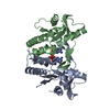
| Entry | Database: PDB / ID: 2o70 | ||||||
|---|---|---|---|---|---|---|---|
| Title | Structure of OHCU decarboxylase from zebrafish | ||||||
 Components Components | OHCU decarboxylase | ||||||
 Keywords Keywords |  LYASE / Uric acid / decarboxylation / 5-hydroxyisourate / allantoin LYASE / Uric acid / decarboxylation / 5-hydroxyisourate / allantoin | ||||||
| Function / homology |  Function and homology information2-oxo-4-hydroxy-4-carboxy-5-ureidoimidazoline decarboxylase activity / 2-oxo-4-hydroxy-4-carboxy-5-ureidoimidazoline decarboxylase / urate catabolic process / allantoin metabolic process / purine nucleobase metabolic process / peroxisome Function and homology information2-oxo-4-hydroxy-4-carboxy-5-ureidoimidazoline decarboxylase activity / 2-oxo-4-hydroxy-4-carboxy-5-ureidoimidazoline decarboxylase / urate catabolic process / allantoin metabolic process / purine nucleobase metabolic process / peroxisomeSimilarity search - Function | ||||||
| Biological species |  Danio rerio (zebrafish) Danio rerio (zebrafish) | ||||||
| Method | X-RAY DIFFRACTION / SYNCHROTRON / MAD / Resolution: 1.8 Å | ||||||
 Authors Authors | Cendron, L. / Berni, R. / Folli, C. / Ramazzina, I. / Percudani, R. / Zanotti, G. | ||||||
 Citation Citation | Journal: J.Biol.Chem. / Year: 2007 Title: The structure of 2-oxo-4-hydroxy-4-carboxy-5-ureidoimidazoline decarboxylase provides insights into the mechanism of uric acid degradation. Authors: Cendron, L. / Berni, R. / Folli, C. / Ramazzina, I. / Percudani, R. / Zanotti, G. | ||||||
| History |
|
- Structure visualization
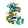
| Structure viewer | Molecule: MolmilJmol/JSmol |
|---|
- Downloads & links
-Download
| PDBx/mmCIF format | 2o70.cif.gz | 227.1 KB | Display | PDBx/mmCIF format |
|---|---|---|---|---|
| PDB format | pdb2o70.ent.gz | 184 KB | Display | PDB format |
| PDBx/mmJSON format | 2o70.json.gz | Tree view | PDBx/mmJSON format | |
| Others |  Other downloads Other downloads |
-Validation report
| Arichive directory | https://data.pdbj.org/pub/pdb/validation_reports/o7/2o70ftp://data.pdbj.org/pub/pdb/validation_reports/o7/2o70 | HTTPS FTP |
|---|
-Related structure data


-Links
 PDBj
PDBj- Assembly
| Deposited unit | 
| ||||||||||||||||||||||||||||||||||||||||||
|---|---|---|---|---|---|---|---|---|---|---|---|---|---|---|---|---|---|---|---|---|---|---|---|---|---|---|---|---|---|---|---|---|---|---|---|---|---|---|---|---|---|---|---|
| 1 | 
| ||||||||||||||||||||||||||||||||||||||||||
| 2 | 
| ||||||||||||||||||||||||||||||||||||||||||
| 3 | 
| ||||||||||||||||||||||||||||||||||||||||||
| Unit cell |
| ||||||||||||||||||||||||||||||||||||||||||
| Noncrystallographic symmetry (NCS) | NCS domain:
NCS domain segments: Component-ID: 1 / Ens-ID: 1 / Beg auth comp-ID: ASP / Beg label comp-ID: ASP / End auth comp-ID: ILE / End label comp-ID: ILE / Refine code: 5 / Auth seq-ID: 2 - 165 / Label seq-ID: 2 - 165
| ||||||||||||||||||||||||||||||||||||||||||
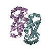
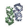
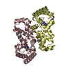
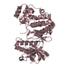
| Details | The biological unit is a dimer. There are three dimers in the asymmetric unit. |
-Components
| #1: Protein | Mass: 19768.645 Da / Num. of mol.: 6 Source method: isolated from a genetically manipulated source Source: (gene. exp.) Danio rerio (zebrafish) / Gene: zgc:158663 / Plasmid: pET28b / Species (production host): Escherichia coli / Production host:  Escherichia coli BL21 (bacteria) / Strain (production host): BL21 / References: UniProt: A1L259 Escherichia coli BL21 (bacteria) / Strain (production host): BL21 / References: UniProt: A1L259#2: Water | ChemComp-HOH / | Water Mass: 18.015 Da / Num. of mol.: 1224 / Source method: isolated from a natural source / Formula: H2O Mass: 18.015 Da / Num. of mol.: 1224 / Source method: isolated from a natural source / Formula: H2O |
|---|
-Experimental details
-Experiment
| Experiment | Method: X-RAY DIFFRACTION / Number of used crystals: 3 |
|---|
- Sample preparation
| Crystal | Density Matthews: 2.48 Å3/Da / Density % sol: 51 % |
|---|---|
| Crystal grow | Temperature: 277 K / Method: vapor diffusion / pH: 8.5 Details: 20 % (v/v) EtOH, 100mM TrisHCl, pH 8.5, VAPOR DIFFUSION, temperature 277K |
-Data collection
| Diffraction |
| |||||||||||||||
|---|---|---|---|---|---|---|---|---|---|---|---|---|---|---|---|---|
| Diffraction source |
| |||||||||||||||
| Detector |
| |||||||||||||||
| Radiation |
| |||||||||||||||
| Radiation wavelength |
| |||||||||||||||
| Reflection | Resolution: 1.8→103.69 Å / Num. all: 111211 / Num. obs: 111211 / % possible obs: 99.9 % / Observed criterion σ(F): 0 / Observed criterion σ(I): 0 / Redundancy: 5.1 % / Rmerge(I) obs: 0.072 / Net I/σ(I): 17.4 | |||||||||||||||
| Reflection shell | Resolution: 1.8→1.9 Å / Redundancy: 4.9 % / Rmerge(I) obs: 0.409 / Mean I/σ(I) obs: 2.8 / Num. unique all: 16284 / % possible all: 100 |

- Processing
| Software |
| ||||||||||||||||||||||||||||||||||||||||||||||||||||||||||||||||||||||||||||||||||||||||||||||||||||||||||||||||||||||||||||||||||||||||||||||||||||||
|---|---|---|---|---|---|---|---|---|---|---|---|---|---|---|---|---|---|---|---|---|---|---|---|---|---|---|---|---|---|---|---|---|---|---|---|---|---|---|---|---|---|---|---|---|---|---|---|---|---|---|---|---|---|---|---|---|---|---|---|---|---|---|---|---|---|---|---|---|---|---|---|---|---|---|---|---|---|---|---|---|---|---|---|---|---|---|---|---|---|---|---|---|---|---|---|---|---|---|---|---|---|---|---|---|---|---|---|---|---|---|---|---|---|---|---|---|---|---|---|---|---|---|---|---|---|---|---|---|---|---|---|---|---|---|---|---|---|---|---|---|---|---|---|---|---|---|---|---|---|---|---|
| Refinement | Method to determine structure: MAD / Resolution: 1.8→67 Å / Cor.coef. Fo:Fc: 0.947 / Cor.coef. Fo:Fc free: 0.928 / SU B: 1.989 / SU ML: 0.064 / Cross valid method: THROUGHOUT / σ(F): 0 / σ(I): 0 / ESU R: 0.13 / ESU R Free: 0.13 / Stereochemistry target values: MAXIMUM LIKELIHOOD / Details: HYDROGENS HAVE BEEN ADDED IN THE RIDING POSITIONS
| ||||||||||||||||||||||||||||||||||||||||||||||||||||||||||||||||||||||||||||||||||||||||||||||||||||||||||||||||||||||||||||||||||||||||||||||||||||||
| Solvent computation | Ion probe radii: 0.8 Å / Shrinkage radii: 0.8 Å / VDW probe radii: 1.4 Å / Solvent model: MASK | ||||||||||||||||||||||||||||||||||||||||||||||||||||||||||||||||||||||||||||||||||||||||||||||||||||||||||||||||||||||||||||||||||||||||||||||||||||||
| Displacement parameters | Biso mean: 22.501 Å2
| ||||||||||||||||||||||||||||||||||||||||||||||||||||||||||||||||||||||||||||||||||||||||||||||||||||||||||||||||||||||||||||||||||||||||||||||||||||||
| Refinement step | Cycle: LAST / Resolution: 1.8→67 Å
| ||||||||||||||||||||||||||||||||||||||||||||||||||||||||||||||||||||||||||||||||||||||||||||||||||||||||||||||||||||||||||||||||||||||||||||||||||||||
| Refine LS restraints |
| ||||||||||||||||||||||||||||||||||||||||||||||||||||||||||||||||||||||||||||||||||||||||||||||||||||||||||||||||||||||||||||||||||||||||||||||||||||||
| Refine LS restraints NCS | Ens-ID: 1 / Refine-ID: X-RAY DIFFRACTION
| ||||||||||||||||||||||||||||||||||||||||||||||||||||||||||||||||||||||||||||||||||||||||||||||||||||||||||||||||||||||||||||||||||||||||||||||||||||||
| LS refinement shell | Resolution: 1.8→1.847 Å / Total num. of bins used: 20
|
