 Movie
Movie Controller


+ Open data

- Basic information
| Entry | Database: PDB / ID: 2o3c | ||||||
|---|---|---|---|---|---|---|---|
| Title | Crystal structure of zebrafish Ape | ||||||
 Components Components | APEX nuclease 1 | ||||||
 Keywords Keywords |  DNA BINDING PROTEIN / APE / ENDONUCLEASE DNA BINDING PROTEIN / APE / ENDONUCLEASE | ||||||
| Function / homology |  Function and homology information Function and homology informationDisplacement of DNA glycosylase by APEX1 / POLB-Dependent Long Patch Base Excision Repair / Resolution of AP sites via the multiple-nucleotide patch replacement pathway / : / Abasic sugar-phosphate removal via the single-nucleotide replacement pathway / Resolution of Abasic Sites (AP sites) / : / phosphodiesterase I activity / : / double-stranded DNA 3'-5' DNA exonuclease activity ...Displacement of DNA glycosylase by APEX1 / POLB-Dependent Long Patch Base Excision Repair / Resolution of AP sites via the multiple-nucleotide patch replacement pathway / : / Abasic sugar-phosphate removal via the single-nucleotide replacement pathway / Resolution of Abasic Sites (AP sites) / : / phosphodiesterase I activity / : / double-stranded DNA 3'-5' DNA exonuclease activity / heart contraction / heart looping / DNA-(apurinic or apyrimidinic site) endonuclease activity / base-excision repair / endonuclease activity / Hydrolases; Acting on ester bonds / nuclear speck / positive regulation of gene expression / nucleolus / negative regulation of apoptotic process / endoplasmic reticulum / mitochondrion / DNA binding / RNA binding / metal ion binding / nucleusSimilarity search - Function | ||||||
| Biological species |  Danio rerio (zebrafish) Danio rerio (zebrafish) | ||||||
| Method | X-RAY DIFFRACTION / SYNCHROTRON / MOLECULAR REPLACEMENT / Resolution: 2.3 Å | ||||||
 Authors Authors | Georgiadis, M.M. / Gaur, R.K. / Delaplane, S. / Svenson, J. | ||||||
 Citation Citation | Journal: Mutat.Res. / Year: 2008 Title: Evolution of the redox function in mammalian apurinic/apyrimidinic endonuclease Authors: Georgiadis, M.M. / Luo, M. / Gaur, R.K. / Delaplane, S. / Li, X. / Kelley, M.R. | ||||||
| History |
|
- Structure visualization
| Structure viewer | Molecule: MolmilJmol/JSmol |
|---|
- Downloads & links
-Download
| PDBx/mmCIF format | 2o3c.cif.gz | 180.7 KB | Display | PDBx/mmCIF format |
|---|---|---|---|---|
| PDB format | pdb2o3c.ent.gz | 143.1 KB | Display | PDB format |
| PDBx/mmJSON format | 2o3c.json.gz | Tree view | PDBx/mmJSON format | |
| Others |  Other downloads Other downloads |
-Validation report
| Arichive directory | https://data.pdbj.org/pub/pdb/validation_reports/o3/2o3cftp://data.pdbj.org/pub/pdb/validation_reports/o3/2o3c | HTTPS FTP |
|---|
-Related structure data
| Related structure data |  2o3hC 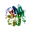 1bixS C: citing same article ( S: Starting model for refinement |
|---|---|
| Similar structure data |


-Links
 PDBj
PDBj


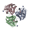
- Assembly
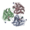
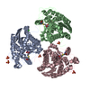
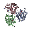
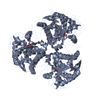
| Deposited unit | 
| ||||||||
|---|---|---|---|---|---|---|---|---|---|
| 1 |
| ||||||||
| Unit cell |
|
-Components
| #1: Protein | Mass: 31952.279 Da / Num. of mol.: 3 Source method: isolated from a genetically manipulated source Source: (gene. exp.) Danio rerio (zebrafish) / Gene: apex1 / Plasmid: PET15B / Production host:  Escherichia coli (E. coli) / Strain (production host): BL21(DE3) ROSETTA / References: UniProt: Q7SXL6, UniProt: A0MTA1*PLUS Escherichia coli (E. coli) / Strain (production host): BL21(DE3) ROSETTA / References: UniProt: Q7SXL6, UniProt: A0MTA1*PLUS#2: Chemical | Lead  Mass: 207.200 Da / Num. of mol.: 3 / Source method: obtained synthetically / Formula: Pb Mass: 207.200 Da / Num. of mol.: 3 / Source method: obtained synthetically / Formula: Pb#3: Water | ChemComp-HOH / | Water Mass: 18.015 Da / Num. of mol.: 362 / Source method: isolated from a natural source / Formula: H2O Mass: 18.015 Da / Num. of mol.: 362 / Source method: isolated from a natural source / Formula: H2O |
|---|
-Experimental details
-Experiment
| Experiment | Method: X-RAY DIFFRACTION / Number of used crystals: 1 |
|---|
- Sample preparation
| Crystal | Density Matthews: 2.8 Å3/Da / Density % sol: 56.7 % |
|---|---|
| Crystal grow | Temperature: 290 K / Method: vapor diffusion, hanging drop / pH: 5.5 Details: AMMONIUM ACETATE, BIS-TRIS, PEG 3350, GLYCEROL, LEAD ACETATE, PH 5.5, pH 5.50, VAPOR DIFFUSION, HANGING DROP, temperature 290K |
-Data collection
| Diffraction | Mean temperature: 100 K |
|---|---|
| Diffraction source | Source: SYNCHROTRON / Site: ALS  / Beamline: 4.2.2 / Wavelength: 0.9 / Beamline: 4.2.2 / Wavelength: 0.9 |
| Detector | Type: NOIR-1 / Detector: CCD / Date: Mar 22, 2005 |
| Radiation | Monochromator: Rosenbaum-Rock monochromator 1: high-resolution double-crystal sagittal focusing, Rosenbaum-Rock monochromator 2: double crystal, Rosenbaum-Rock vertical focusing mirror Protocol: SINGLE WAVELENGTH / Monochromatic (M) / Laue (L): M / Scattering type: x-ray |
| Radiation wavelength | Wavelength: 0.9 Å / Relative weight: 1 |
| Reflection | Resolution: 2.3→50 Å / Num. all: 47731 / Num. obs: 46075 / % possible obs: 96.5 % / Redundancy: 3.07 % / Biso Wilson estimate: 30.1 Å2 / Rmerge(I) obs: 0.059 / Net I/σ(I): 14.3 |
| Reflection shell | Resolution: 2.3→2.4 Å / Redundancy: 2.94 % / Rmerge(I) obs: 0.268 / Mean I/σ(I) obs: 3.4 / % possible all: 85.5 |
- Processing
| Software |
| ||||||||||||||||||||||||||||
|---|---|---|---|---|---|---|---|---|---|---|---|---|---|---|---|---|---|---|---|---|---|---|---|---|---|---|---|---|---|
| Refinement | Method to determine structure: MOLECULAR REPLACEMENT Starting model: PDB ENTRY 1BIX Resolution: 2.3→50 Å / Isotropic thermal model: ISOTROPIC / Cross valid method: THROUGHOUT / σ(F): 0 / Stereochemistry target values: Engh & Huber / Details: MAXIMUM LIKELIHOOD
| ||||||||||||||||||||||||||||
| Displacement parameters | Biso mean: 28.3 Å2
| ||||||||||||||||||||||||||||
| Refine analyze |
| ||||||||||||||||||||||||||||
| Refinement step | Cycle: LAST / Resolution: 2.3→50 Å
| ||||||||||||||||||||||||||||
| Refine LS restraints |
| ||||||||||||||||||||||||||||
| LS refinement shell | Resolution: 2.3→2.4 Å
|
