 Movie
Movie Controller

[English] 日本語
 Yorodumi
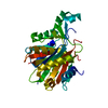
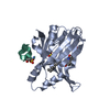
- PDB-2o0m: The crystal structure of the putative SorC family transcriptional... -
+ Open data

- Basic information
| Entry | Database: PDB / ID: 2o0m | ||||||
|---|---|---|---|---|---|---|---|
| Title | The crystal structure of the putative SorC family transcriptional regulator from Enterococcus faecalis | ||||||
 Components Components | Transcriptional regulator, SorC family Transcriptional regulation Transcriptional regulation | ||||||
 Keywords Keywords | TRANSCRIPTION / SorC family / transcriptional regulator / Structural Genomics / PSI-2 / Protein Structure Initiative / Midwest Center for Structural Genomics / MCSG | ||||||
| Function / homology |  Function and homology information Function and homology information | ||||||
| Biological species |   Enterococcus faecalis (bacteria) Enterococcus faecalis (bacteria) | ||||||
| Method | X-RAY DIFFRACTION / SYNCHROTRON / SAD / Resolution: 1.6 Å | ||||||
 Authors Authors | Zhang, R. / Zhou, M. / Bargassa, M. / Joachimiak, A. / Midwest Center for Structural Genomics (MCSG) | ||||||
 Citation Citation | Journal: To be Published Title: The crystal structure of the putative SorC family transcriptional regulator from Enterococcus faecalis Authors: Zhang, R. / Zhou, M. / Bargassa, M. / Joachimiak, A. | ||||||
| History |
| ||||||
| Remark 300 | BIOMOLECULE: 1 THIS ENTRY CONTAINS THE CRYSTALLOGRAPHIC ASYMMETRIC UNIT WHICH CONSISTS OF 1 CHAIN(S) ...BIOMOLECULE: 1 THIS ENTRY CONTAINS THE CRYSTALLOGRAPHIC ASYMMETRIC UNIT WHICH CONSISTS OF 1 CHAIN(S). THE BIOLOGICAL UNIT OF THE PROTEIN IS NOT KNOWN |
- Structure visualization
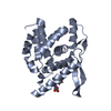
| Structure viewer | Molecule: MolmilJmol/JSmol |
|---|
- Downloads & links
-Download
| PDBx/mmCIF format | 2o0m.cif.gz | 124.7 KB | Display | PDBx/mmCIF format |
|---|---|---|---|---|
| PDB format | pdb2o0m.ent.gz | 95.8 KB | Display | PDB format |
| PDBx/mmJSON format | 2o0m.json.gz | Tree view | PDBx/mmJSON format | |
| Others |  Other downloads Other downloads |
-Validation report
| Arichive directory | https://data.pdbj.org/pub/pdb/validation_reports/o0/2o0mftp://data.pdbj.org/pub/pdb/validation_reports/o0/2o0m | HTTPS FTP |
|---|
-Related structure data
| Similar structure data | |
|---|---|
| Other databases |


-Links
 PDBj
PDBj- Assembly
| Deposited unit | 
| ||||||||||||
|---|---|---|---|---|---|---|---|---|---|---|---|---|---|
| 1 |
| ||||||||||||
| Unit cell |
| ||||||||||||
| Components on special symmetry positions |
|
-Components
| #1: Protein | Transcriptional regulation Mass: 39391.637 Da / Num. of mol.: 1 Source method: isolated from a genetically manipulated source Source: (gene. exp.) Enterococcus faecalis (bacteria) / Strain: V583 / Plasmid: pET15b / Species (production host): Escherichia coli / Production host: Escherichia coli BL21(DE3) (bacteria) / Strain (production host): BL21(DE3) / References: UniProt: Q833I7 | ||
|---|---|---|---|
| #2: Chemical | Phosphate  Mass: 94.971 Da / Num. of mol.: 2 / Source method: obtained synthetically / Formula: PO4 Mass: 94.971 Da / Num. of mol.: 2 / Source method: obtained synthetically / Formula: PO4#3: Water | ChemComp-HOH / | Water Mass: 18.015 Da / Num. of mol.: 377 / Source method: isolated from a natural source / Formula: H2O Mass: 18.015 Da / Num. of mol.: 377 / Source method: isolated from a natural source / Formula: H2O |
-Experimental details
-Experiment
| Experiment | Method: X-RAY DIFFRACTION / Number of used crystals: 1 |
|---|
- Sample preparation
| Crystal | Density Matthews: 1.72 Å3/Da |
|---|---|
| Crystal grow | Temperature: 298 K / Method: vapor diffusion, sitting drop / pH: 8.5 Details: 0.2M Ammonium Sulfate, 0.1M Tris, 25% PEG3350, pH 8.5, VAPOR DIFFUSION, SITTING DROP, temperature 298K |
-Data collection
| Diffraction | Mean temperature: 100 K |
|---|---|
| Diffraction source | Source: SYNCHROTRON / Site: APS  / Beamline: 19-ID / Wavelength: 0.9798 Å / Beamline: 19-ID / Wavelength: 0.9798 Å |
| Detector | Type: ADSC QUANTUM 315 / Detector: CCD / Date: Feb 25, 2006 / Details: mirrors |
| Radiation | Monochromator: Si 111 channel / Protocol: SINGLE WAVELENGTH / Monochromatic (M) / Laue (L): M / Scattering type: x-ray |
| Radiation wavelength | Wavelength: 0.9798 Å / Relative weight: 1 |
| Reflection | Resolution: 1.6→56.52 Å / Num. all: 33906 / Num. obs: 33635 / % possible obs: 0.2 % / Observed criterion σ(F): 2 / Observed criterion σ(I): 2 / Redundancy: 4.6 % / Biso Wilson estimate: 16 Å2 / Rmerge(I) obs: 0.096 / Net I/σ(I): 15.93 |
| Reflection shell | Resolution: 1.6→1.642 Å / Redundancy: 4.1 % / Rmerge(I) obs: 0.291 / Mean I/σ(I) obs: 2.46 / Num. unique all: 2468 / % possible all: 93.48 |
- Processing
| Software |
| |||||||||||||||||||||||||||||||||||||||||||||||||||||||||||||||||||||||||||||||||||||||||||||||||||||||||||||||||||||||||||||
|---|---|---|---|---|---|---|---|---|---|---|---|---|---|---|---|---|---|---|---|---|---|---|---|---|---|---|---|---|---|---|---|---|---|---|---|---|---|---|---|---|---|---|---|---|---|---|---|---|---|---|---|---|---|---|---|---|---|---|---|---|---|---|---|---|---|---|---|---|---|---|---|---|---|---|---|---|---|---|---|---|---|---|---|---|---|---|---|---|---|---|---|---|---|---|---|---|---|---|---|---|---|---|---|---|---|---|---|---|---|---|---|---|---|---|---|---|---|---|---|---|---|---|---|---|---|---|
| Refinement | Method to determine structure: SAD / Resolution: 1.6→56.52 Å / Cor.coef. Fo:Fc: 0.965 / Cor.coef. Fo:Fc free: 0.945 / SU B: 3.546 / SU ML: 0.058 / Cross valid method: THROUGHOUT / σ(F): 0 / σ(I): 0 / ESU R: 0.112 / ESU R Free: 0.086 Stereochemistry target values: MAXIMUM LIKELIHOOD WITH PHASES Details: HYDROGENS HAVE BEEN ADDED IN THE RIDING POSITIONS
| |||||||||||||||||||||||||||||||||||||||||||||||||||||||||||||||||||||||||||||||||||||||||||||||||||||||||||||||||||||||||||||
| Solvent computation | Ion probe radii: 0.8 Å / Shrinkage radii: 0.8 Å / VDW probe radii: 1.2 Å / Solvent model: MASK | |||||||||||||||||||||||||||||||||||||||||||||||||||||||||||||||||||||||||||||||||||||||||||||||||||||||||||||||||||||||||||||
| Displacement parameters | Biso mean: 15.952 Å2
| |||||||||||||||||||||||||||||||||||||||||||||||||||||||||||||||||||||||||||||||||||||||||||||||||||||||||||||||||||||||||||||
| Refine analyze |
| |||||||||||||||||||||||||||||||||||||||||||||||||||||||||||||||||||||||||||||||||||||||||||||||||||||||||||||||||||||||||||||
| Refinement step | Cycle: LAST / Resolution: 1.6→56.52 Å
| |||||||||||||||||||||||||||||||||||||||||||||||||||||||||||||||||||||||||||||||||||||||||||||||||||||||||||||||||||||||||||||
| Refine LS restraints |
| |||||||||||||||||||||||||||||||||||||||||||||||||||||||||||||||||||||||||||||||||||||||||||||||||||||||||||||||||||||||||||||
| LS refinement shell | Resolution: 1.6→1.642 Å / Total num. of bins used: 20
| |||||||||||||||||||||||||||||||||||||||||||||||||||||||||||||||||||||||||||||||||||||||||||||||||||||||||||||||||||||||||||||
| Refinement TLS params. | Method: refined / Origin x: -0.866 Å / Origin y: 26.651 Å / Origin z: 16.703 Å
| |||||||||||||||||||||||||||||||||||||||||||||||||||||||||||||||||||||||||||||||||||||||||||||||||||||||||||||||||||||||||||||
| Refinement TLS group |
|
