 Movie
Movie Controller

[English] 日本語
 Yorodumi
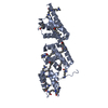
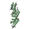
- PDB-2nrj: Crystal Structure of Hemolysin binding component from Bacillus cereus -
+ Open data

- Basic information
| Entry | Database: PDB / ID: 2nrj | ||||||
|---|---|---|---|---|---|---|---|
| Title | Crystal Structure of Hemolysin binding component from Bacillus cereus | ||||||
 Components Components | Hbl B protein | ||||||
 Keywords Keywords |  TOXIN / Enterotoxin / Hemolysis / Transmembrane / Structural Genomics / PSI-2 / Protein Structure Initiative / New York SGX Research Center for Structural Genomics / NYSGXRC TOXIN / Enterotoxin / Hemolysis / Transmembrane / Structural Genomics / PSI-2 / Protein Structure Initiative / New York SGX Research Center for Structural Genomics / NYSGXRC | ||||||
| Function / homology |  Function and homology informationhemolysis in another organism / : / toxin activity / membrane => GO:0016020 / host cell plasma membrane / extracellular region / membrane Function and homology informationhemolysis in another organism / : / toxin activity / membrane => GO:0016020 / host cell plasma membrane / extracellular region / membraneSimilarity search - Function | ||||||
| Biological species |  Bacillus cereus (bacteria) Bacillus cereus (bacteria) | ||||||
| Method | X-RAY DIFFRACTION / SYNCHROTRON / SAD / Resolution: 2.03 Å | ||||||
 Authors Authors | Madegowda, M. / Eswaramoorthy, S. / Burley, S.K. / Swaminathan, S. / New York SGX Research Center for Structural Genomics (NYSGXRC) | ||||||
 Citation Citation | Journal: Proteins / Year: 2008 Title: X-ray crystal structure of the B component of Hemolysin BL from Bacillus cereus Authors: Madegowda, M. / Eswaramoorthy, S. / Burley, S.K. / Swaminathan, S. | ||||||
| History |
|
- Structure visualization
| Structure viewer | Molecule: MolmilJmol/JSmol |
|---|
- Downloads & links
-Download
| PDBx/mmCIF format | 2nrj.cif.gz | 82.1 KB | Display | PDBx/mmCIF format |
|---|---|---|---|---|
| PDB format | pdb2nrj.ent.gz | 61.7 KB | Display | PDB format |
| PDBx/mmJSON format | 2nrj.json.gz | Tree view | PDBx/mmJSON format | |
| Others |  Other downloads Other downloads |
-Validation report
| Arichive directory | https://data.pdbj.org/pub/pdb/validation_reports/nr/2nrjftp://data.pdbj.org/pub/pdb/validation_reports/nr/2nrj | HTTPS FTP |
|---|
-Related structure data
| Similar structure data | |
|---|---|
| Other databases |


-Links
 PDBj
PDBj- Assembly
| Deposited unit | 
| |||||||||
|---|---|---|---|---|---|---|---|---|---|---|
| 1 |
| |||||||||
| Unit cell |
| |||||||||
| Components on special symmetry positions |
|
-Components
| #1: Protein | Mass: 38934.703 Da / Num. of mol.: 1 / Fragment: residues 32-375 Source method: isolated from a genetically manipulated source Source: (gene. exp.) Bacillus cereus (bacteria) / Gene: hblA / Plasmid: pSGX4 (BS) / Production host: Escherichia coli (E. coli) / Strain (production host): Bl21(DE3)+Codon+RIL / References: UniProt: Q9REG6, UniProt: P80172*PLUS |
|---|---|
| #2: Water | ChemComp-HOH / Water Mass: 18.015 Da / Num. of mol.: 199 / Source method: isolated from a natural source / Formula: H2O Mass: 18.015 Da / Num. of mol.: 199 / Source method: isolated from a natural source / Formula: H2O |
-Experimental details
-Experiment
| Experiment | Method: X-RAY DIFFRACTION / Number of used crystals: 1 |
|---|
- Sample preparation
| Crystal | Density Matthews: 2.03 Å3/Da / Density % sol: 39.43 % |
|---|---|
| Crystal grow | Temperature: 298 K / Method: vapor diffusion, sitting drop / pH: 8.5 Details: 30% PEG 4000, 0.2M Magnesium Chloride, 0.1M Tris, pH 8.5, VAPOR DIFFUSION, SITTING DROP, temperature 298.0K |
-Data collection
| Diffraction | Mean temperature: 100 K |
|---|---|
| Diffraction source | Source: SYNCHROTRON / Site: NSLS  / Beamline: X12C / Wavelength: 0.9772 Å / Beamline: X12C / Wavelength: 0.9772 Å |
| Detector | Type: ADSC QUANTUM 210 / Detector: CCD / Date: Oct 30, 2006 / Details: mirrors |
| Radiation | Monochromator: Si(111) / Protocol: SINGLE WAVELENGTH / Monochromatic (M) / Laue (L): M / Scattering type: x-ray |
| Radiation wavelength | Wavelength: 0.9772 Å / Relative weight: 1 |
| Reflection | Resolution: 2.03→26.05 Å / Num. all: 20797 / Num. obs: 20797 / % possible obs: 99.7 % / Observed criterion σ(F): 0 / Observed criterion σ(I): 0 / Redundancy: 7.2 % / Biso Wilson estimate: 12.4 Å2 / Rmerge(I) obs: 0.085 / Net I/σ(I): 17.5 |
| Reflection shell | Resolution: 2.03→2.1 Å / Redundancy: 6.1 % / Rmerge(I) obs: 0.272 / Num. unique all: 1974 / % possible all: 97 |
- Processing
| Software |
| ||||||||||||||||||||||||||||||||||||||||||||||||||||||||||||||||||||||||||||||||
|---|---|---|---|---|---|---|---|---|---|---|---|---|---|---|---|---|---|---|---|---|---|---|---|---|---|---|---|---|---|---|---|---|---|---|---|---|---|---|---|---|---|---|---|---|---|---|---|---|---|---|---|---|---|---|---|---|---|---|---|---|---|---|---|---|---|---|---|---|---|---|---|---|---|---|---|---|---|---|---|---|---|
| Refinement | Method to determine structure: SAD / Resolution: 2.03→26.05 Å / Rfactor Rfree error: 0.008 / Data cutoff high absF: 92972.21 / Data cutoff low absF: 0 / Isotropic thermal model: RESTRAINED / Cross valid method: THROUGHOUT / σ(F): 0 / Stereochemistry target values: Engh & Huber Details: The authors state that the missing residues listed in remark 465 are due to lack of electron density
| ||||||||||||||||||||||||||||||||||||||||||||||||||||||||||||||||||||||||||||||||
| Solvent computation | Solvent model: FLAT MODEL / Bsol: 40.8406 Å2 / ksol: 0.368405 e/Å3 | ||||||||||||||||||||||||||||||||||||||||||||||||||||||||||||||||||||||||||||||||
| Displacement parameters | Biso mean: 20.7 Å2
| ||||||||||||||||||||||||||||||||||||||||||||||||||||||||||||||||||||||||||||||||
| Refine analyze |
| ||||||||||||||||||||||||||||||||||||||||||||||||||||||||||||||||||||||||||||||||
| Refinement step | Cycle: LAST / Resolution: 2.03→26.05 Å
| ||||||||||||||||||||||||||||||||||||||||||||||||||||||||||||||||||||||||||||||||
| Refine LS restraints |
| ||||||||||||||||||||||||||||||||||||||||||||||||||||||||||||||||||||||||||||||||
| LS refinement shell | Resolution: 2.03→2.16 Å / Rfactor Rfree error: 0.026 / Total num. of bins used: 6
| ||||||||||||||||||||||||||||||||||||||||||||||||||||||||||||||||||||||||||||||||
| Xplor file |
|
