 Movie
Movie Controller


+ Open data

- Basic information
| Entry | Database: PDB / ID: 2nqn | ||||||
|---|---|---|---|---|---|---|---|
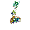
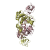
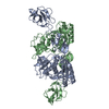
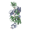
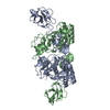
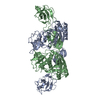
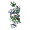
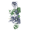
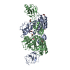
| Title | MoeA T100W | ||||||
 Components Components | Molybdopterin biosynthesis protein moeA | ||||||
 Keywords Keywords |  BIOSYNTHETIC PROTEIN / molybdopterin / MPT / Moco / molybdenum / MoeA / MogA / gephyrin / Cnx1 / cinnamon BIOSYNTHETIC PROTEIN / molybdopterin / MPT / Moco / molybdenum / MoeA / MogA / gephyrin / Cnx1 / cinnamon | ||||||
| Function / homology |  Function and homology informationmolybdopterin molybdotransferase / molybdopterin molybdotransferase activity / Mo-molybdopterin cofactor biosynthetic process / protein homodimerization activity / identical protein binding / metal ion binding / cytosol / cytoplasm Function and homology informationmolybdopterin molybdotransferase / molybdopterin molybdotransferase activity / Mo-molybdopterin cofactor biosynthetic process / protein homodimerization activity / identical protein binding / metal ion binding / cytosol / cytoplasmSimilarity search - Function | ||||||
| Biological species |  Escherichia coli (E. coli) Escherichia coli (E. coli) | ||||||
| Method | X-RAY DIFFRACTION / SYNCHROTRON / MOLECULAR REPLACEMENT / Resolution: 2.2 Å | ||||||
 Authors Authors | Nicolas, J. / Xiang, S. / Schindelin, H. / Rajagopalan, K.V. | ||||||
 Citation Citation | Journal: Biochemistry / Year: 2007 Title: Mutational Analysis of Escherichia coli MoeA: Two Functional Activities Map to the Active Site Cleft. Authors: Nichols, J.D. / Xiang, S. / Schindelin, H. / Rajagopalan, K.V. | ||||||
| History |
|
- Structure visualization
| Structure viewer | Molecule: MolmilJmol/JSmol |
|---|
- Downloads & links
-Download
| PDBx/mmCIF format | 2nqn.cif.gz | 177.7 KB | Display | PDBx/mmCIF format |
|---|---|---|---|---|
| PDB format | pdb2nqn.ent.gz | 142.1 KB | Display | PDB format |
| PDBx/mmJSON format | 2nqn.json.gz | Tree view | PDBx/mmJSON format | |
| Others |  Other downloads Other downloads |
-Validation report
| Arichive directory | https://data.pdbj.org/pub/pdb/validation_reports/nq/2nqnftp://data.pdbj.org/pub/pdb/validation_reports/nq/2nqn | HTTPS FTP |
|---|
-Related structure data
| Related structure data |  2nqkC  2nqmC  2nqqC 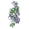 2nqrC  2nqsC 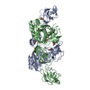 2nquC  2nqvC  2nroC 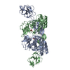 2nrpC  2nrsC C: citing same article ( |
|---|---|
| Similar structure data |


-Links
 PDBj
PDBj- Assembly
| Deposited unit | 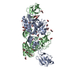
| ||||||||||||||||||||||||||||||||||||||||||||||||||||||||||||||||||||||||||||||||||||||||||||||||||
|---|---|---|---|---|---|---|---|---|---|---|---|---|---|---|---|---|---|---|---|---|---|---|---|---|---|---|---|---|---|---|---|---|---|---|---|---|---|---|---|---|---|---|---|---|---|---|---|---|---|---|---|---|---|---|---|---|---|---|---|---|---|---|---|---|---|---|---|---|---|---|---|---|---|---|---|---|---|---|---|---|---|---|---|---|---|---|---|---|---|---|---|---|---|---|---|---|---|---|---|
| 1 |
| ||||||||||||||||||||||||||||||||||||||||||||||||||||||||||||||||||||||||||||||||||||||||||||||||||
| Unit cell |
| ||||||||||||||||||||||||||||||||||||||||||||||||||||||||||||||||||||||||||||||||||||||||||||||||||
| Noncrystallographic symmetry (NCS) | NCS domain:
NCS domain segments: Ens-ID: 1
| ||||||||||||||||||||||||||||||||||||||||||||||||||||||||||||||||||||||||||||||||||||||||||||||||||
| Details | Dimer of chain A and B |
-Components
| #1: Protein | Mass: 44191.164 Da / Num. of mol.: 2 / Mutation: T100W Source method: isolated from a genetically manipulated source Source: (gene. exp.) Escherichia coli (E. coli) / Gene: moeA, bisB, chlE, narE / Plasmid: pET11a / Production host: Escherichia coli (E. coli) / Strain (production host): Ah69 (DE3) / References: UniProt: P12281#2: Chemical | ChemComp-GOL / Glycerol  Mass: 92.094 Da / Num. of mol.: 29 / Source method: obtained synthetically / Formula: C3H8O3 Mass: 92.094 Da / Num. of mol.: 29 / Source method: obtained synthetically / Formula: C3H8O3#3: Water | ChemComp-HOH / | Water Mass: 18.015 Da / Num. of mol.: 474 / Source method: isolated from a natural source / Formula: H2O Mass: 18.015 Da / Num. of mol.: 474 / Source method: isolated from a natural source / Formula: H2O |
|---|
-Experimental details
-Experiment
| Experiment | Method: X-RAY DIFFRACTION / Number of used crystals: 1 |
|---|
- Sample preparation
| Crystal | Density Matthews: 3.06 Å3/Da / Density % sol: 59.85 % |
|---|---|
| Crystal grow | Temperature: 298 K / Method: vapor diffusion, hanging drop / pH: 6.5 Details: 10%-15% PEG 6000/8000, 0.2-0.5 M calcium acetate, 0.1 M cacodylic acid, pH 6.5, VAPOR DIFFUSION, HANGING DROP, temperature 298K |
-Data collection
| Diffraction | Mean temperature: 100 K |
|---|---|
| Diffraction source | Source: SYNCHROTRON / Site: NSLS  / Beamline: X26C / Wavelength: 1.1 / Beamline: X26C / Wavelength: 1.1 |
| Detector | Type: ADSC QUANTUM 4 / Detector: CCD / Date: Apr 16, 2001 |
| Radiation | Protocol: SINGLE WAVELENGTH / Monochromatic (M) / Laue (L): M / Scattering type: x-ray |
| Radiation wavelength | Wavelength: 1.1 Å / Relative weight: 1 |
| Reflection | Resolution: 2.2→50 Å / Num. all: 51582 / Num. obs: 51582 / % possible obs: 100 % / Observed criterion σ(F): 2 / Observed criterion σ(I): 2 |
- Processing
| Software |
| ||||||||||||||||||||||||||||||||||||||||||||||||||||||||||||||||||||||||||||||||||||||||||||||||||||
|---|---|---|---|---|---|---|---|---|---|---|---|---|---|---|---|---|---|---|---|---|---|---|---|---|---|---|---|---|---|---|---|---|---|---|---|---|---|---|---|---|---|---|---|---|---|---|---|---|---|---|---|---|---|---|---|---|---|---|---|---|---|---|---|---|---|---|---|---|---|---|---|---|---|---|---|---|---|---|---|---|---|---|---|---|---|---|---|---|---|---|---|---|---|---|---|---|---|---|---|---|---|
| Refinement | Method to determine structure: MOLECULAR REPLACEMENT / Resolution: 2.2→49.39 Å / Cor.coef. Fo:Fc: 0.943 / Cor.coef. Fo:Fc free: 0.915 / Cross valid method: THROUGHOUT / σ(F): 0 / ESU R: 0.264 / ESU R Free: 0.222 / Stereochemistry target values: MAXIMUM LIKELIHOOD
| ||||||||||||||||||||||||||||||||||||||||||||||||||||||||||||||||||||||||||||||||||||||||||||||||||||
| Solvent computation | Ion probe radii: 0.8 Å / Shrinkage radii: 0.8 Å / VDW probe radii: 1.4 Å / Solvent model: BABINET MODEL WITH MASK | ||||||||||||||||||||||||||||||||||||||||||||||||||||||||||||||||||||||||||||||||||||||||||||||||||||
| Displacement parameters | Biso mean: 42.951 Å2
| ||||||||||||||||||||||||||||||||||||||||||||||||||||||||||||||||||||||||||||||||||||||||||||||||||||
| Refinement step | Cycle: LAST / Resolution: 2.2→49.39 Å
| ||||||||||||||||||||||||||||||||||||||||||||||||||||||||||||||||||||||||||||||||||||||||||||||||||||
| Refine LS restraints |
| ||||||||||||||||||||||||||||||||||||||||||||||||||||||||||||||||||||||||||||||||||||||||||||||||||||
| Refine LS restraints NCS | Dom-ID: 1 / Auth asym-ID: A / Ens-ID: 1 / Refine-ID: X-RAY DIFFRACTION
| ||||||||||||||||||||||||||||||||||||||||||||||||||||||||||||||||||||||||||||||||||||||||||||||||||||
| LS refinement shell | Resolution: 2.2→2.257 Å / Total num. of bins used: 20
|
