 Movie
Movie Controller

[English] 日本語
 Yorodumi
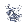
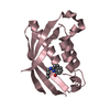
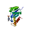
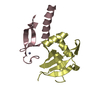
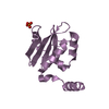
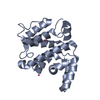
- PDB-2n3t: Solution structure of the Rpn1 substrate receptor site toroid 1 (T1) -
+ Open data

- Basic information
| Entry | Database: PDB / ID: 2n3t | ||||||
|---|---|---|---|---|---|---|---|
| Title | Solution structure of the Rpn1 substrate receptor site toroid 1 (T1) | ||||||
 Components Components | 26S proteasome regulatory subunit RPN1 | ||||||
 Keywords Keywords |  PROTEIN BINDING PROTEIN BINDING | ||||||
| Function / homology |  Function and homology information Function and homology informationproteasome regulatory particle, base subcomplex / Cross-presentation of soluble exogenous antigens (endosomes) / TNFR2 non-canonical NF-kB pathway / regulation of protein catabolic process / proteasome storage granule / Ub-specific processing proteases / enzyme regulator activity / Neutrophil degranulation / proteasome complex / protein-macromolecule adaptor activity ...proteasome regulatory particle, base subcomplex / Cross-presentation of soluble exogenous antigens (endosomes) / TNFR2 non-canonical NF-kB pathway / regulation of protein catabolic process / proteasome storage granule / Ub-specific processing proteases / enzyme regulator activity / Neutrophil degranulation / proteasome complex / protein-macromolecule adaptor activity / ubiquitin-dependent protein catabolic process / proteasome-mediated ubiquitin-dependent protein catabolic process / endoplasmic reticulum / nucleus / cytoplasmSimilarity search - Function | ||||||
| Biological species |  Saccharomyces cerevisiae (brewer's yeast) Saccharomyces cerevisiae (brewer's yeast) | ||||||
| Method | SOLUTION NMR / simulated annealing | ||||||
 Authors Authors | Chen, X. / Walters, K.J. | ||||||
 Citation Citation | Journal: Science / Year: 2016 Title: Rpn1 provides adjacent receptor sites for substrate binding and deubiquitination by the proteasome. Authors: Shi, Y. / Chen, X. / Elsasser, S. / Stocks, B.B. / Tian, G. / Lee, B.H. / Shi, Y. / Zhang, N. / de Poot, S.A. / Tuebing, F. / Sun, S. / Vannoy, J. / Tarasov, S.G. / Engen, J.R. / Finley, D. / Walters, K.J. | ||||||
| History |
|
- Structure visualization
| Structure viewer | Molecule: MolmilJmol/JSmol |
|---|
- Downloads & links
-Download
| PDBx/mmCIF format | 2n3t.cif.gz | 381 KB | Display | PDBx/mmCIF format |
|---|---|---|---|---|
| PDB format | pdb2n3t.ent.gz | 314.2 KB | Display | PDB format |
| PDBx/mmJSON format | 2n3t.json.gz | Tree view | PDBx/mmJSON format | |
| Others |  Other downloads Other downloads |
-Validation report
| Arichive directory | https://data.pdbj.org/pub/pdb/validation_reports/n3/2n3tftp://data.pdbj.org/pub/pdb/validation_reports/n3/2n3t | HTTPS FTP |
|---|
-Related structure data
| Related structure data |  2n3uC  2n3vC 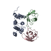 2n3wC C: citing same article ( |
|---|---|
| Similar structure data | |
| Other databases |


-Links
 PDBj
PDBj


- Assembly
| Deposited unit | 
| |||||||||
|---|---|---|---|---|---|---|---|---|---|---|
| 1 |
| |||||||||
| NMR ensembles |
|
-Components
| #1: Protein | Mass: 13636.592 Da / Num. of mol.: 1 / Fragment: UNP residues 482-612 Source method: isolated from a genetically manipulated source Source: (gene. exp.) Saccharomyces cerevisiae (brewer's yeast)Strain: ATCC 204508 / S288c / Gene: RPN1, HRD2, NAS1, RPD1, YHR027C / Plasmid: Pgex4t1 / Production host:  Escherichia coli (E. coli) / References: UniProt: P38764 Escherichia coli (E. coli) / References: UniProt: P38764 |
|---|
-Experimental details
-Experiment
| Experiment | Method: SOLUTION NMR | ||||||||||||||||||||||||||||||||||||||||
|---|---|---|---|---|---|---|---|---|---|---|---|---|---|---|---|---|---|---|---|---|---|---|---|---|---|---|---|---|---|---|---|---|---|---|---|---|---|---|---|---|---|
| NMR experiment |
|
- Sample preparation
| Details |
| ||||||||||||||||||||||||||||||||||||||||||||||||||||||||||||||||||||||||||||
|---|---|---|---|---|---|---|---|---|---|---|---|---|---|---|---|---|---|---|---|---|---|---|---|---|---|---|---|---|---|---|---|---|---|---|---|---|---|---|---|---|---|---|---|---|---|---|---|---|---|---|---|---|---|---|---|---|---|---|---|---|---|---|---|---|---|---|---|---|---|---|---|---|---|---|---|---|---|
| Sample |
| ||||||||||||||||||||||||||||||||||||||||||||||||||||||||||||||||||||||||||||
| Sample conditions | Ionic strength: 50 / pH: 6.7 / Pressure: ambient / Temperature: 298 K |
-NMR measurement
| NMR spectrometer |
|
|---|
- Processing
| NMR software |
| |||||||||||||||||||||||||||
|---|---|---|---|---|---|---|---|---|---|---|---|---|---|---|---|---|---|---|---|---|---|---|---|---|---|---|---|---|
| Refinement | Method: simulated annealing / Software ordinal: 1 | |||||||||||||||||||||||||||
| NMR constraints | NOE constraints total: 1242 / NOE intraresidue total count: 576 / NOE long range total count: 247 / NOE medium range total count: 241 / NOE sequential total count: 178 / Protein phi angle constraints total count: 91 / Protein psi angle constraints total count: 91 | |||||||||||||||||||||||||||
| NMR representative | Selection criteria: lowest energy | |||||||||||||||||||||||||||
| NMR ensemble | Conformer selection criteria: structures with the lowest energy Conformers calculated total number: 50 / Conformers submitted total number: 10 / Maximum lower distance constraint violation: 1.8 Å / Maximum upper distance constraint violation: 5.5 Å |
