 Movie
Movie Controller

[English] 日本語
 Yorodumi
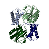
- PDB-2mjp: STRUCTURE-BASED IDENTIFICATION OF THE BIOCHEMICAL FUNCTION OF A H... -
+ Open data

- Basic information
| Entry | Database: PDB / ID: 2mjp | ||||||
|---|---|---|---|---|---|---|---|
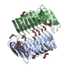
| Title | STRUCTURE-BASED IDENTIFICATION OF THE BIOCHEMICAL FUNCTION OF A HYPOTHETICAL PROTEIN FROM METHANOCOCCUS JANNASCHII:MJ0226 | ||||||
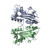
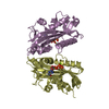
 Components Components | PYROPHOSPHATASE | ||||||
 Keywords Keywords | STRUCTURAL GENOMICS / PYROPHOSPHATASE / HYPERTHERMAL PROTEIN / BSGC structure funded by NIH / Protein Structure Initiative / PSI / Berkeley Structural Genomics Center | ||||||
| Function / homology |  Function and homology information Function and homology informationXTP/dITP diphosphatase / purine nucleoside triphosphate catabolic process / ITP diphosphatase activity / XTP diphosphatase activity / dITP diphosphatase activity / nucleoside triphosphate catabolic process / nucleoside triphosphate diphosphatase activity / nucleotide metabolic process / ribonucleoside triphosphate phosphatase activity / nucleotide binding ...XTP/dITP diphosphatase / purine nucleoside triphosphate catabolic process / ITP diphosphatase activity / XTP diphosphatase activity / dITP diphosphatase activity / nucleoside triphosphate catabolic process / nucleoside triphosphate diphosphatase activity / nucleotide metabolic process / ribonucleoside triphosphate phosphatase activity / nucleotide binding / metal ion binding / cytosol / cytoplasmSimilarity search - Function | ||||||
| Biological species |   Methanocaldococcus jannaschii (archaea) Methanocaldococcus jannaschii (archaea) | ||||||
| Method | X-RAY DIFFRACTION / MOLECULAR REPLACEMENT / Resolution: 2.2 Å | ||||||
 Authors Authors | Hwang, K.Y. / Chung, J.H. / Han, Y.S. / Kim, S.H. / Cho, Y. / Berkeley Structural Genomics Center (BSGC) | ||||||
 Citation Citation | Journal: Nat.Struct.Biol. / Year: 1999 Title: Structure-based identification of a novel NTPase from Methanococcus jannaschii. Authors: Hwang, K.Y. / Chung, J.H. / Kim, S.H. / Han, Y.S. / Cho, Y. | ||||||
| History |
|
- Structure visualization
| Structure viewer | Molecule: MolmilJmol/JSmol |
|---|
- Downloads & links
-Download
| PDBx/mmCIF format | 2mjp.cif.gz | 88.7 KB | Display | PDBx/mmCIF format |
|---|---|---|---|---|
| PDB format | pdb2mjp.ent.gz | 67.6 KB | Display | PDB format |
| PDBx/mmJSON format | 2mjp.json.gz | Tree view | PDBx/mmJSON format | |
| Others |  Other downloads Other downloads |
-Validation report
| Arichive directory | https://data.pdbj.org/pub/pdb/validation_reports/mj/2mjpftp://data.pdbj.org/pub/pdb/validation_reports/mj/2mjp | HTTPS FTP |
|---|
-Related structure data


-Links
 PDBj
PDBj


- Assembly
| Deposited unit | 
| ||||||||
|---|---|---|---|---|---|---|---|---|---|
| 1 |
| ||||||||
| Unit cell |
|
-Components
| #1: Protein | Mass: 22232.453 Da / Num. of mol.: 2 / Source method: isolated from a natural source / Details: AMPPNP COMPLEX STRUCTURE / Source: (natural) Methanocaldococcus jannaschii (archaea) / References: UniProt: Q57679#2: Chemical | ChemComp-ANP / |   Mass: 506.196 Da / Num. of mol.: 1 / Source method: obtained synthetically / Formula: C10H17N6O12P3 / Comment: AMP-PNP, energy-carrying molecule analogue*YM Mass: 506.196 Da / Num. of mol.: 1 / Source method: obtained synthetically / Formula: C10H17N6O12P3 / Comment: AMP-PNP, energy-carrying molecule analogue*YM#3: Water | ChemComp-HOH / | Water Mass: 18.015 Da / Num. of mol.: 161 / Source method: isolated from a natural source / Formula: H2O Mass: 18.015 Da / Num. of mol.: 161 / Source method: isolated from a natural source / Formula: H2O |
|---|
-Experimental details
-Experiment
| Experiment | Method: X-RAY DIFFRACTION / Number of used crystals: 1 |
|---|
- Sample preparation
| Crystal | Density Matthews: 2.53 Å3/Da / Density % sol: 51.34 % | ||||||||||||||||||||||||||||||||||||||||||
|---|---|---|---|---|---|---|---|---|---|---|---|---|---|---|---|---|---|---|---|---|---|---|---|---|---|---|---|---|---|---|---|---|---|---|---|---|---|---|---|---|---|---|---|
| Crystal grow | pH: 6.5 / Details: pH 6.5 | ||||||||||||||||||||||||||||||||||||||||||
| Crystal grow | *PLUS pH: 7.5 / Method: vapor diffusion | ||||||||||||||||||||||||||||||||||||||||||
| Components of the solutions | *PLUS
|
-Data collection
| Diffraction | Mean temperature: 100 K |
|---|---|
| Diffraction source | Wavelength: 1.542 |
| Radiation | Protocol: SINGLE WAVELENGTH / Monochromatic (M) / Laue (L): M / Scattering type: x-ray |
| Radiation wavelength | Wavelength: 1.542 Å / Relative weight: 1 |
| Reflection | Resolution: 2.2→50 Å / Num. obs: 20346 / % possible obs: 93 % / Redundancy: 3.8 % / Biso Wilson estimate: 8.5 Å2 / Rmerge(I) obs: 0.049 / Rsym value: 6.6 |
- Processing
| Software | Name: X-PLOR / Version: 3.851 / Classification: refinement | ||||||||||||||||||||||||||||||||||||||||||||||||||||||||||||
|---|---|---|---|---|---|---|---|---|---|---|---|---|---|---|---|---|---|---|---|---|---|---|---|---|---|---|---|---|---|---|---|---|---|---|---|---|---|---|---|---|---|---|---|---|---|---|---|---|---|---|---|---|---|---|---|---|---|---|---|---|---|
| Refinement | Method to determine structure: MOLECULAR REPLACEMENT / Resolution: 2.2→8 Å / Rfactor Rfree error: 0.006 / Cross valid method: THROUGHOUT / σ(F): 2
| ||||||||||||||||||||||||||||||||||||||||||||||||||||||||||||
| Displacement parameters | Biso mean: 16.9 Å2 | ||||||||||||||||||||||||||||||||||||||||||||||||||||||||||||
| Refine analyze |
| ||||||||||||||||||||||||||||||||||||||||||||||||||||||||||||
| Refinement step | Cycle: LAST / Resolution: 2.2→8 Å
| ||||||||||||||||||||||||||||||||||||||||||||||||||||||||||||
| Refine LS restraints |
| ||||||||||||||||||||||||||||||||||||||||||||||||||||||||||||
| LS refinement shell | Resolution: 2.2→2.33 Å / Rfactor Rfree error: 0.017 / Total num. of bins used: 6
| ||||||||||||||||||||||||||||||||||||||||||||||||||||||||||||
| Xplor file |
| ||||||||||||||||||||||||||||||||||||||||||||||||||||||||||||
| Software | *PLUS Version: 3.851 / Classification: refinement | ||||||||||||||||||||||||||||||||||||||||||||||||||||||||||||
| Refine LS restraints | *PLUS
|
