 Movie
Movie Controller

[English] 日本語
 Yorodumi
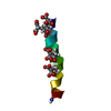
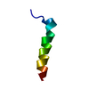
- PDB-2mcv: Solid-state NMR structure of piscidin 1 in aligned 1:1 phosphatid... -
+ Open data

- Basic information
| Entry | Database: PDB / ID: 2mcv | ||||||
|---|---|---|---|---|---|---|---|
| Title | Solid-state NMR structure of piscidin 1 in aligned 1:1 phosphatidylethanolamine/phosphoglycerol lipid bilayers | ||||||
 Components Components | Moronecidin | ||||||
 Keywords Keywords |  ANTIMICROBIAL PROTEIN / antimicrobial peptide / anticancer peptide / anti HIV-1 / cationic / amphipathic / histidine rich / helical / lipid bilayers / bacterial cell membrane mimic ANTIMICROBIAL PROTEIN / antimicrobial peptide / anticancer peptide / anti HIV-1 / cationic / amphipathic / histidine rich / helical / lipid bilayers / bacterial cell membrane mimic | ||||||
| Function / homology | Pleurocidin / Pleurocidin family / defense response to fungus / killing of cells of another organism / defense response to bacterium / extracellular region / Moronecidin Function and homology information Function and homology information | ||||||
| Biological species |  Morone saxatilis (striped sea-bass) Morone saxatilis (striped sea-bass) | ||||||
| Method | SOLID-STATE NMR / simulated annealing | ||||||
| Model details | lowest energy, model1 | ||||||
 Authors Authors | Fu, R. / Tian, Y. / Perrin Jr., B.S. / Grant, C.V. / Hayden, R.M. / Pastor, R.W. / Cotten, M.L. | ||||||
 Citation Citation | Journal: J.Am.Chem.Soc. / Year: 2014 Title: High-resolution structures and orientations of antimicrobial peptides piscidin 1 and piscidin 3 in fluid bilayers reveal tilting, kinking, and bilayer immersion. Authors: Perrin, B.S. / Tian, Y. / Fu, R. / Grant, C.V. / Chekmenev, E.Y. / Wieczorek, W.E. / Dao, A.E. / Hayden, R.M. / Burzynski, C.M. / Venable, R.M. / Sharma, M. / Opella, S.J. / Pastor, R.W. / Cotten, M.L. | ||||||
| History |
|
- Structure visualization
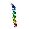
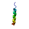
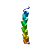
| Structure viewer | Molecule: MolmilJmol/JSmol |
|---|
- Downloads & links
-Download
| PDBx/mmCIF format | 2mcv.cif.gz | 75.9 KB | Display | PDBx/mmCIF format |
|---|---|---|---|---|
| PDB format | pdb2mcv.ent.gz | 54.6 KB | Display | PDB format |
| PDBx/mmJSON format | 2mcv.json.gz | Tree view | PDBx/mmJSON format | |
| Others |  Other downloads Other downloads |
-Validation report
| Arichive directory | https://data.pdbj.org/pub/pdb/validation_reports/mc/2mcvftp://data.pdbj.org/pub/pdb/validation_reports/mc/2mcv | HTTPS FTP |
|---|
-Related structure data
| Related structure data |  2mcuC  2mcwC  2mcxC C: citing same article ( |
|---|---|
| Similar structure data | |
| Other databases |


-Links
 PDBj
PDBj- Assembly
| Deposited unit | 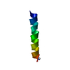
| |||||||||
|---|---|---|---|---|---|---|---|---|---|---|
| 1 |
| |||||||||
| NMR ensembles |
|
-Components
| #1: Protein/peptide | Mass: 2577.085 Da / Num. of mol.: 1 / Source method: obtained synthetically / Details: Synthetic construct / Source: (synth.) Morone saxatilis (striped sea-bass) / References: UniProt: Q8UUG0 |
|---|
-Experimental details
-Experiment
| Experiment | Method: SOLID-STATE NMR |
|---|---|
| NMR experiment | Type: 15N 1H solid-state de-HETCOR |
- Sample preparation
| Details | Contents: 15-20 MM PISCIDIN 1, 300-400 MM 1:1 (MOLAR) 1-PALMITOYL-2-OLEOYL-SN-GLYCERO-PHOSPHATIDYLETHANOLAMINE/1-PALMITOYL-2-OLEOYL-SN-GLYCERO-PHOSPHOGLYCEROL, 40 MM PHOSPHATE BUFFER WITH 100% H2O Solvent system: 100% H2O |
|---|---|
| Sample conditions | pH: 6.0 / Pressure: ambient / Temperature: 305 K |
-NMR measurement
| NMR spectrometer |
|
|---|
- Processing
| NMR software |
| |||||||||
|---|---|---|---|---|---|---|---|---|---|---|
| Refinement | Method: simulated annealing / Software ordinal: 1 Details: Structures were calculated using a simulated annealing protocol within Xplor-NIH with torsion angle molecular dynamics in the presence of experimentally determined restraints. Solid-state ...Details: Structures were calculated using a simulated annealing protocol within Xplor-NIH with torsion angle molecular dynamics in the presence of experimentally determined restraints. Solid-state NMR experiments on static oriented lipid bilayer samples allowed for the measurements of anisotropic backbone 15N chemical shifts and 15N-1H dipolar couplings, which were used as the experimental restraints. The initial structure was an alpha helix with ideal phi/psi angles (-61/-45). The calculations also included the Xplor-NIH potential for knowledge-based torsion angles and the routine terms ANGL, BOND and IMPR. A total of 100 structures were generated and the 10 lowest energy structures were accepted for analysis and representation. By convention, the bilayer normal for all of the oriented samples is aligned along the z-axis of the calculated structures. | |||||||||
| NMR representative | Selection criteria: lowest energy | |||||||||
| NMR ensemble | Conformer selection criteria: structures with the lowest energy Conformers calculated total number: 100 / Conformers submitted total number: 10 |
