 Movie
Movie Controller

[English] 日本語
 Yorodumi
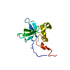
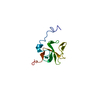
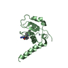
- PDB-2khi: NMR structure of the domain 4 of the E. coli ribosomal protein S1 -
+ Open data

- Basic information
| Entry | Database: PDB / ID: 2khi | ||||||
|---|---|---|---|---|---|---|---|
| Title | NMR structure of the domain 4 of the E. coli ribosomal protein S1 | ||||||
 Components Components | 30S ribosomal protein S1 | ||||||
 Keywords Keywords | RIBOSOMAL PROTEIN / ribosomal protein S1 / Acetylation / Phosphoprotein / Ribonucleoprotein / RNA-binding | ||||||
| Function / homology |  Function and homology information Function and homology informationRNA secondary structure unwinding / positive regulation of cytoplasmic translation / negative regulation of cytoplasmic translation / ribosomal small subunit assembly / cytosolic small ribosomal subunit / cytoplasmic translation / single-stranded RNA binding / structural constituent of ribosome / translation / mRNA binding ...RNA secondary structure unwinding / positive regulation of cytoplasmic translation / negative regulation of cytoplasmic translation / ribosomal small subunit assembly / cytosolic small ribosomal subunit / cytoplasmic translation / single-stranded RNA binding / structural constituent of ribosome / translation / mRNA binding / RNA binding / membrane / cytoplasmSimilarity search - Function | ||||||
| Biological species |  Escherichia coli (E. coli) Escherichia coli (E. coli) | ||||||
| Method | SOLUTION NMR / torsion angle dynamics, simulated annealing | ||||||
 Authors Authors | Salah, P. / Bisaglia, M. / Aliprandi, P. / Uzan, M. / Sizun, C. / Bontems, F. | ||||||
 Citation Citation | Journal: Nucleic Acids Res. / Year: 2009 Title: Probing the relationship between Gram-negative and Gram-positive S1 proteins by sequence analysis Authors: Salah, P. / Bisaglia, M. / Aliprandi, P. / Uzan, M. / Sizun, C. / Bontems, F. | ||||||
| History |
|
- Structure visualization
| Structure viewer | Molecule: MolmilJmol/JSmol |
|---|
- Downloads & links
-Download
| PDBx/mmCIF format | 2khi.cif.gz | 361.9 KB | Display | PDBx/mmCIF format |
|---|---|---|---|---|
| PDB format | pdb2khi.ent.gz | 296.6 KB | Display | PDB format |
| PDBx/mmJSON format | 2khi.json.gz | Tree view | PDBx/mmJSON format | |
| Others |  Other downloads Other downloads |
-Validation report
| Arichive directory | https://data.pdbj.org/pub/pdb/validation_reports/kh/2khiftp://data.pdbj.org/pub/pdb/validation_reports/kh/2khi | HTTPS FTP |
|---|
-Related structure data


-Links
 PDBj
PDBj


- Assembly
| Deposited unit | 
| |||||||||
|---|---|---|---|---|---|---|---|---|---|---|
| 1 |
| |||||||||
| NMR ensembles |
|
-Components
| #1: Protein | Mass: 13010.658 Da / Num. of mol.: 1 / Fragment: UNP residues 267-361 Source method: isolated from a genetically manipulated source Source: (gene. exp.) Escherichia coli (E. coli) / Gene: rpsA, ssyF, b0911, JW0894 / Plasmid: pET15b / Production host: Escherichia coli (E. coli) / Strain (production host): BL21(DE3) / References: UniProt: P0AG67 |
|---|
-Experimental details
-Experiment
| Experiment | Method: SOLUTION NMR | ||||||||||||||||
|---|---|---|---|---|---|---|---|---|---|---|---|---|---|---|---|---|---|
| NMR experiment |
|
- Sample preparation
| Details |
| ||||||||||||||||||||
|---|---|---|---|---|---|---|---|---|---|---|---|---|---|---|---|---|---|---|---|---|---|
| Sample |
| ||||||||||||||||||||
| Sample conditions | Ionic strength: 200 / pH: 6.8 / Pressure: ambient / Temperature: 303 K |
-NMR measurement
| NMR spectrometer | Type: Bruker DRX / Manufacturer: Bruker / Model: DRX / Field strength: 600 MHz |
|---|
- Processing
| NMR software |
| |||||||||
|---|---|---|---|---|---|---|---|---|---|---|
| Refinement | Method: torsion angle dynamics, simulated annealing / Software ordinal: 1 | |||||||||
| NMR ensemble | Conformer selection criteria: 12 structures for lowest energy Conformers calculated total number: 100 / Conformers submitted total number: 12 / Representative conformer: 1 |
